Review
. 2012 Feb;135(Pt 2):320-44.
doi: 10.1093/brain/awr271. Epub 2011 Nov 8.

Neurological diseases and pain

Affiliations
Review

Neurological diseases and pain

David Borsook. Brain. 2012 Feb.

Abstract

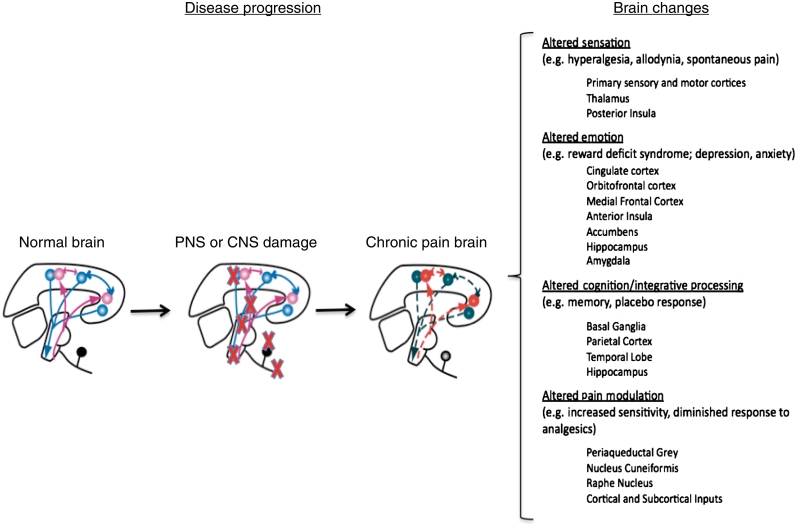
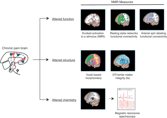
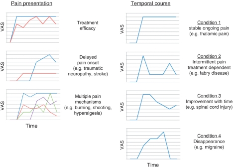
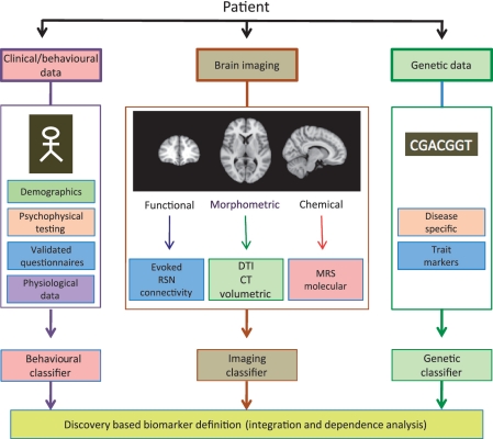
Chronic pain is a frequent component of many neurological disorders, affecting 20-40% of patients for many primary neurological diseases. These diseases result from a wide range of pathophysiologies including traumatic injury to the central nervous system, neurodegeneration and neuroinflammation, and exploring the aetiology of pain in these disorders is an opportunity to achieve new insight into pain processing. Whether pain originates in the central or peripheral nervous system, it frequently becomes centralized through maladaptive responses within the central nervous system that can profoundly alter brain systems and thereby behaviour (e.g. depression). Chronic pain should thus be considered a brain disease in which alterations in neural networks affect multiple aspects of brain function, structure and chemistry. The study and treatment of this disease is greatly complicated by the lack of objective measures for either the symptoms or the underlying mechanisms of chronic pain. In pain associated with neurological disease, it is sometimes difficult to obtain even a subjective evaluation of pain, as is the case for patients in a vegetative state or end-stage Alzheimer's disease. It is critical that neurologists become more involved in chronic pain treatment and research (already significant in the fields of migraine and peripheral neuropathies). To achieve this goal, greater efforts are needed to enhance training for neurologists in pain treatment and promote greater interest in the field. This review describes examples of pain in different neurological diseases including primary neurological pain conditions, discusses the therapeutic potential of brain-targeted therapies and highlights the need for objective measures of pain.


Figures

Figure 1
Figure 1
Brain changes in chronic pain. The figure summarizes two concepts that relate to the development of chronic pain following damage to either peripheral or CNS pathways involved in pain. (i) The first is that following injury (red crosses) progressive changes take place in the brain: the normal brain is altered in a manner that produces changes in function and structure in the chronic pain state; (ii) the second, noted in the text on the right, indicates that multiple brain regions involved in sensation, emotion, cognition and pain modulation may manifest in varied behavioural symptoms from ongoing pain to anxiety and depression. While it is easy to conceptualize altered systems, altered circuitry is best considered in the context of interactive brain processes that are disrupted in chronic pain. Both central and peripheral origins of pain are noted; the peripheral sources are clearly important in producing or maintaining central changes whether these are part of a disease affecting both central and peripheral systems or not.
Figure 2
Figure 2
Changes in function, structure and chemistry in chronic pain. Chronic pain alters the brain (left) and produces alterations in function [e.g. increased activation as in central sensitization (Lee et al., 2008)]; altered resting state networks (Apkarian et al., 2004); and altered chemistry [e.g. changes in excitatory and inhibitory amino acids, (Gussew et al., 2011)]. While the example of nuclear magnetic resonance (NMR) approaches for evaluating alterations in the brain in pain conditions (Borsook et al., 2007; Borsook and Becerra, 2011) is provided, other approaches have been employed to measure pain-related alterations in brain systems including electroencephalography (Brinkmeyer et al., 2010), magnetoencephalography (Maihofner et al., 2010) and near infrared spectroscopy (Slater et al., 2006; Becerra et al., 2008). fa = fractional anisotropy; fMRI = functional MRI.
Figure 3
Figure 3
Chronic pain symptoms and variations in temporal course. Chronobiological effects of pain, treatment effects, environmental changes (e.g. barometric pressure) activity all may contribute to a variation in pain over time (Auvil-Novak, 1999; Lake, 2005; Odrcich et al., 2006; Dworkin et al., 2007; Kloss-Brandstatter et al., 2011). Left: Examples of features of pain presentation. (i) Relatively small effect of therapy (red line) versus pain (blue line) [pain is rated by patients on an 11-point (0–10) Likert's scale (VAS)] usually decreases by just two points on this scale; on average most pharmacological treatments decrease pain by 20–30% in population placebo controlled studies; (ii) the onset of pain may be variable and even be delayed following disease onset (red line)—this is more easily observed with specific PNS or CNS damage (i.e. traumatic neuropathy or thalamic stroke); (iii) most conditions have more than one pain (lines in different colours representing different pains) that can be defined mechanistically—e.g. shooting pain due to ectopic activity; burning pain due to inhibitory neuron dropout (Baron et al., 2010). Right: Representative examples of different temporal courses for pain from constant (Condition 1) to intermittent or dependent on specific treatment (Condition 2) to a natural course of improvement (Condition 3) to complete remission (Condition 4). The colours are used to differentiate time courses where multiple plots are used.
Figure 4
Figure 4
Objective imaging assays. Current research for pain biomarkers includes brain imaging (Borsook et al., 2011a, b), clinical/behavioural approaches (Freynhagen et al., 2006; Scholz et al., 2009) and genetic markers (Tegeder et al., 2006). Each of these approaches has yielded some promising results, suggesting that specific classifiers can be defined that may be used alone or, more likely, in combination, as pain biomarkers. DTI = diffusion tensor imaging; RSN = resting state network; MRS = magnetic resonance spectroscopy.
Figure 5
Figure 5
Schematic of altered pain processing in neurological disease. The figure summarizes altered pain processing in examples of neurological disease, as well as underlying mechanisms that contribute to chronic disease-related pain. In most diseases, multiple regions are affected (as opposed to secondary effects such as centralization of pain following a peripheral nerve injury). Whether or not the disease is a primary or secondary cause of pain, pain itself drives an altered brain state (Fig. 1). Damage anywhere along the pain pathway from peripheral nerve to spinothalamic tracts and more central pathways including thalamus and thalamocortical projections may result in neuropathic pain. In the figure, damage as indicated by red crosses or abnormal activation in pain pathways (circles), contributes to other changes in brain systems.

References

    1. Abbruzzese G, Berardelli A. Sensorimotor integration in movement disorders. Mov Disord. 2003;18:231–40. - PubMed
    1. Aghakhani N, Parker F, David P, Morar S, Lacroix C, Benoudiba F, et al. Long-term follow-up of Chiari-related syringomyelia in adults: analysis of 157 surgically treated cases. Neurosurgery. 2009;64:308–15. - PubMed
    1. Albano B, Dinia L, Del Sette M, Gandolfo C, Sivori G, Finocchi C. Fabry disease in patients with migraine with aura. Neurol Sci. 2010;31(Suppl 1):S167–9. - PubMed
    1. Albin RL, Young AB. Somatosensory phenomena in Huntington's disease. Mov Disord. 1988;3:343–6. - PubMed
    1. Alderfer BS, Arciniegas DB, Silver JM. Treatment of depression following traumatic brain injury. J Head Trauma Rehabil. 2005;20:544–62. - PubMed

Publication types

MeSH terms