. 2012 Feb;14(2):132-44.
doi: 10.1093/neuonc/nor195. Epub 2011 Nov 7.

Maintenance of primary tumor phenotype and genotype in glioblastoma stem cells

Affiliations

Maintenance of primary tumor phenotype and genotype in glioblastoma stem cells

Hiroaki Wakimoto et al. Neuro Oncol. 2012 Feb.

Abstract

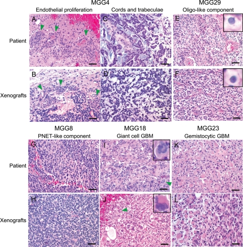
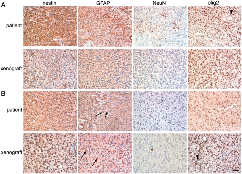
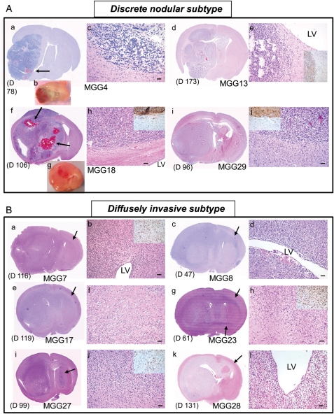
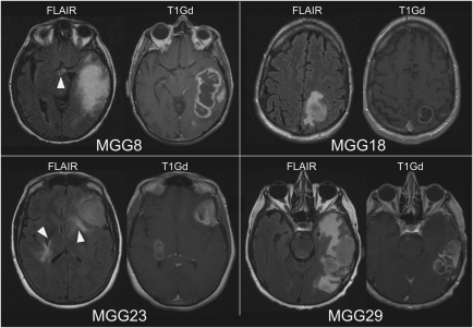
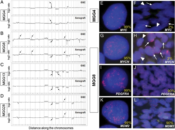
The clinicopathological heterogeneity of glioblastoma (GBM) and the various genetic and phenotypic subtypes in GBM stem cells (GSCs) are well described. However, the relationship between GSCs and the corresponding primary tumor from which they were isolated is poorly understood. We have established GSC-enriched neurosphere cultures from 15 newly diagnosed GBM specimens and examined the relationship between the histopathological and genomic features of GSC-derived orthotopic xenografts and those of the respective patient tumors. GSC-initiated xenografts recapitulate the distinctive cytological hallmarks and diverse histological variants associated with the corresponding patient GBM, including giant cell and gemistocytic GBM, and primitive neuroectodermal tumor (PNET)-like components. This indicates that GSCs generate tumors that preserve patient-specific disease phenotypes. The majority of GSC-derived intracerebral xenografts (11 of 15) demonstrated a highly invasive behavior crossing the midline, whereas the remainder formed discrete nodular and vascular masses. In some cases, GSC invasiveness correlated with preoperative MRI, but not with the status of PI3-kinase/Akt pathways or O(6)-methylguanine methyltransferase expression. Genome-wide screening by array comparative genomic hybridization and fluorescence in situ hybridization revealed that GSCs harbor unique genetic copy number aberrations. GSCs acquiring amplifications of the myc family genes represent only a minority of tumor cells within the original patient tumors. Thus, GSCs are a genetically distinct subpopulation of neoplastic cells within a GBM. These studies highlight the value of GSCs for preclinical modeling of clinically relevant, patient-specific GBM and, thus, pave the way for testing novel anti-GSC/GBM agents for personalized therapy.


Figures

Fig. 1.
Fig. 1.
GSC-derived xenografts recapitulate histopathological features of the original patient GBM. Top and third rows, primary tumors from patients; second and bottom rows, intracerebral xenografts derived from GSCs. (A and B) MGG4 showing endothelial proliferation (arrowheads). (C and D) Tumor cells arranged in cords and trabeculae in MGG4. (E and F) Oligodendroglial component observed in MGG29 was recapitulated in its respective xenografts. Insets, cells with perinuclear halo. (G and H) Many MGG8 tumor cells display undifferentiated cytological features like primitive neuroectodermal tumors (PNETs). (I and J) MGG18 tumor is characterized by marked cellular pleomorphism and giant, bizarre cells, features of giant cell GBM. Arrowheads and insets show multinucleated giant cells. (K and L) MGG23 contains tumor cells with abundant eosinophilic cytoplasm indicative of the gemistocytic phenotype. Bars, 50 µm.
Fig. 2.
Fig. 2.
Immunohistochemical comparison between the original tumor and GSC-derived intracerebral xenografts. (A) MGG28 and (B) MGG23. Upper panels, primary patient tumor; Lower panels, intracerebral xenograft generated by GSCs. From left to right: staining for neural progenitor marker nestin, astrocytic marker GFAP, neuronal marker NeuN, glioma/oligodendroglial/stem cell marker olig2. Arrows show GFAP-positive gemistocytic cells present in both patient and GSC MGG23 tumors. Note negative staining of olig2 in blood vessels (arrowheads). Bar, 50 µm.
Fig. 3.
Fig. 3.
Invasiveness subtyping of GSC-derived GBM xenografts. (A) Discrete nodular subtype. a-c, MGG4. d, e, MGG13. f-h, MGG18. i, j, MGG29. H & E staining of coronal brain sections, except b and g, which show coronally cut planes of freshly removed brains. These tumors are characterized by relatively delineated borders between tumor mass and brain parenchyma, which was confirmed by immunohistochemistry for human-specific nestin (insets in e, h, and j). This tumor subtype was associated with increased vascularity (b, g) and intratumoral bleeding (arrows in a and f). There is typically no tendency for cells to accumulate in the subventricular zones (adjacent LV) (e, h). (B) Diffusely invasive subtype. a, b, MGG7. c, d, MGG8. e, f, MGG17. g, h, MGG23. i, j, MGG27. k, l, MGG28. These tumors are diffuse and highly invasive, and always extend to the contralateral hemisphere through white matter tracts (shown by arrows). Indistinct tumor-brain interface (f, h, and j; insets in b, h, and j showing human-specific nestin-positive cells on infiltrative edge) and heavy neoplastic infiltration in the subventricular zone (b, d, and l) are other phenotypic characteristics of this subtype of tumor. The timing of animal sacrifice when tumors caused significant symptoms is shown in days. (D) For each panel. LV, lateral ventricle. Bars, 50 µm.
Fig. 4.
Fig. 4.
Patient preoperative MRIs. MR images taken before surgeries showing MGG8 and MGG23 that produced GSCs of highly invasive phenotype, and MGG18 and MGG29, from which GSCs of discrete nodular phenotype were derived. Arrowheads show hyperintense abnormal lesions in FLAIR images at the midline or bilateral regions suggestive of tumor infiltration. T1Gd, T1 images after infusion of gadolinium contrast agent.
Fig. 5.
Fig. 5.
Genomic profiles of GSCs and identification of GSC-associated gene copy abnormalities in primary tumors. (A–D) aCGH analysis reveals gene copy number aberrations in cultured GSCs (upper panels) and xenografts derived from GSCs (lower panels). (A) MGG4, arrow shows amplification of CMYC on 8q24.2. (B) MGG8, arrows show amplifications of MYCN (2p24.3), PDGFRA (4q12), MDM2 (12q15), and homozygous deletion of CDKN2A/B (9p21.3) genes. (C) MGG13, arrows show homozygous deletion of CDKN2A/B genes (9p21.3) and 10q23.31 locus. (D) MGG28, arrows show homozygous deletion of CDKN2A/B (9p21.3) and amplification of CDK4 (12q14) genes. (E–L) FISH analysis of MYC in MGG4 GSCs. (E) and primary tumor (F), and MYCN (G), PDGFRA (I) and MDM2 (K) in MGG8 GSCs and primary tumors (H, J, L). Amplifications are shown by red spots in nuclei. Green spots indicate hybridization with a control probe. In F and H, arrows indicate cells with, and arrowheads without MYC/MYCN amplification. Numbers in yellow denote the percentage of gene-amplified cells in GSCs and primary tumors.

References

    1. Wen PY, Kesari S. Malignant gliomas in adults. N Engl J Med. 2008;359:492–507. doi:10.1056/NEJMra0708126. - DOI - PubMed
    1. Stupp R, Hegi ME, Mason WP, et al. Effects of radiotherapy with concomitant and adjuvant temozolomide versus radiotherapy alone on survival in glioblastoma in a randomised phase III study: 5-year analysis of the EORTC-NCIC trial. Lancet Oncol. 2009;10:459–466. doi:10.1016/S1470-2045(09)70025-7. - DOI - PubMed
    1. Stupp R, Mason WP, van den Bent MJ, et al. Radiotherapy plus concomitant and adjuvant temozolomide for glioblastoma. N Engl J Med. 2005;352:987–996. doi:10.1056/NEJMoa043330. - DOI - PubMed
    1. Miller CR, Perry A. Glioblastoma. Arch Pathol Lab Med. 2007;131:397–406. - PubMed
    1. Comprehensive genomic characterization defines human glioblastoma genes and core pathways. Nature. 2008;455:1061–1068. doi:10.1038/nature07385. - DOI - PMC - PubMed

Publication types

MeSH terms

Substances