. 2012 Jan 18;31(2):471-80.
doi: 10.1038/emboj.2011.402. Epub 2011 Nov 8.

An indirect role for ASPP1 in limiting p53-dependent p21 expression and cellular senescence

Affiliations

An indirect role for ASPP1 in limiting p53-dependent p21 expression and cellular senescence

Arnaud M Vigneron et al. EMBO J. .

Abstract

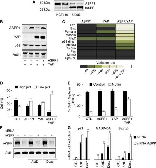
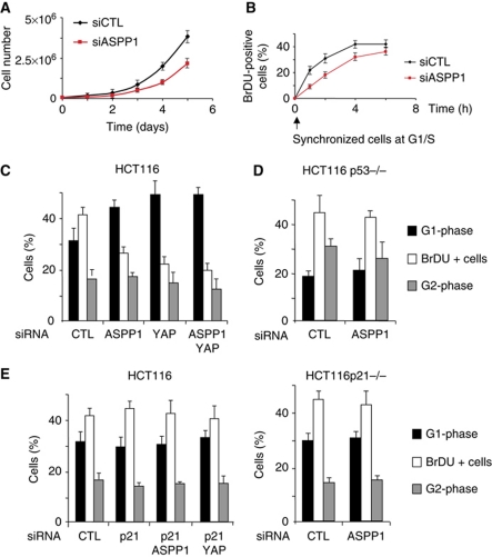
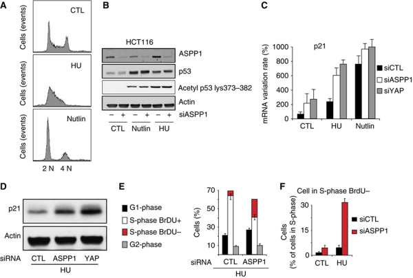
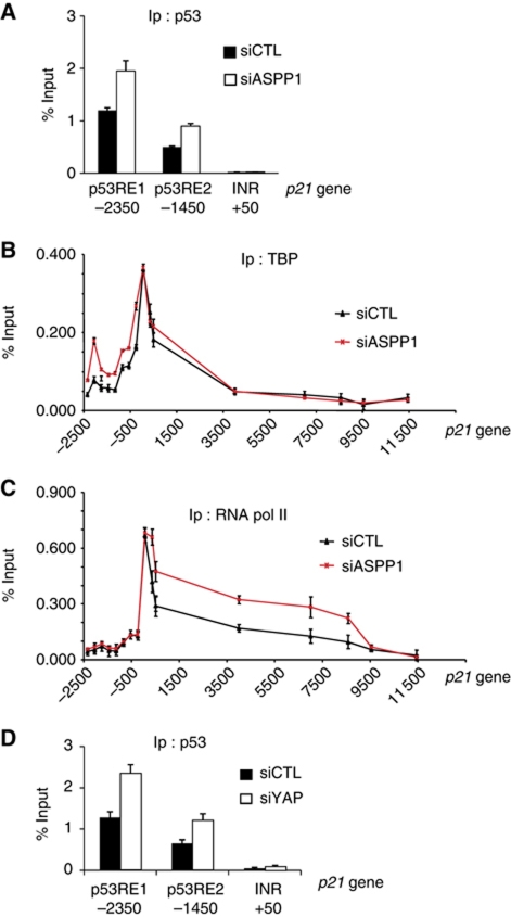
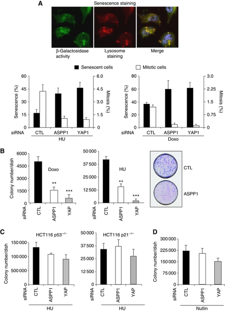
In addition to acting as a transcriptional cofactor for p53, ASPP1 has been shown to function in the cytoplasm to regulate the nuclear localization and activity of YAP/TAZ. We show here that the ability of ASPP1 to activate YAP results in the decreased expression of LATS2, which lowers the ability of p53 to induce p21, cell-cycle arrest and senescence. ASPP1 expression peaks in S-phase, and down-regulation of ASPP1 leads to a reduction in DNA synthesis and enhanced senescence in response to drugs that impede DNA replication. These activities of cytoplasmic ASPP1 in opposing p53-mediated p21 expression are in contrast to the role of nuclear ASPP1 in cooperating with p53 to induce the expression of apoptotic target genes, and may help to dampen p53 activity in normal cells.


Conflict of interest statement

The authors declare that they have no conflict of interest.

Figures

Figure 1
Figure 1
ASPP1/YAP overexpression inhibits p53 transcriptional activity. (A) ASPP1 and iASPP expression detected by western blot with specific antibodies in HCT116 and U2OS cells. Duplicate samples from each cell line are shown. (B) U2OS cells were infected with retroviral constructs coding for ASPP1 and YAP as shown and expression of p53, ASPP1 and YAP detected by western blot 2 days later. Actin expression was used as a loading control. (C) U2OS cells were infected as in (B) and mRNA expression of different p53-target genes tested by RT–qPCR using specific primers. The results were normalized against two different standard genes, and the graphs represent the mean of three independent experiments. (D) U2OS cells were transfected with vectors coding for ASPP1 and YAP as indicated and treated for 24 h with 10 μM Nutlin. ASPP1 and p21 expression was detected by immunofluorescence with specific antibodies. In the absence of treatment, p21 staining was extremely low in all cells—following treatment cells staining brightly for p21 were scored as showing high expression while cells staining at background levels were scored as showing low expression. (E) Cells infected as in (B) and treated with Nutlin as in (D) were analysed for BrdU incorporation and DNA content analysis by flow cytometry. (F) U2OS cells were transfected with control siRNA or specific siRNAs directed against iASPP. iASPP expression was detected by western blot 2 days later. Actin was used as a loading control. (G) U2OS cells were transfected as in (F) and mRNA expression of indicated p53-target genes was tested by RT–qPCR using specific primers after treatment with 5 nM of Actinomycin D (ActD) or 200 ng/ml of Doxorubicin (Doxo) for 24 h. The results were normalized against two different standard genes, and the graphs represent the mean of three independent experiments.
Figure 2
Figure 2
Inhibition of HCT116 cell proliferation after ASPP1/YAP down-regulation is p53 and p21 dependent. (A) Cell proliferation of HCT116 cells transfected with control or ASPP1 siRNA was quantified by Trypan blue exclusion. (B) Cells transfected as in (A) were synchronized at the G1/S-phase transition by double thymidine block, and analysed for BrdU incorporation at the indicated times following release. (C) Cell cycle of HCT116 transfected by the indicated siRNA was analysed by BrdU and PI incorporation, measured by flow cytometry. Results represent the mean of three independent experiments. (D) p53−/− HCT116 cells were transfected and analysed as in (C). Results represent the mean of three independent experiments. (E) HCT116 cells transfected with siRNA targeting p21 (LHS), or p21−/− cells (RHS) were treated and analysed as in (C). Results represent the mean of three independent experiments. Note the control siRNA data in (E) are the same as those shown in (C).
Figure 3
Figure 3
Increase of the p53 response to DNA replication inhibition following ASPP1 and YAP down-regulation. (A) Cell cycle of HCT116 treated for 24 h with 400 μM of HU or 10 μM of Nutlin, analysed by BrdU and PI incorporation and measured by flow cytometry. Result shows a typical histogram for the different conditions. (B) HCT116 cells transfected by control siRNA or siRNA against ASPP1 and treated as in (A) were analysed by western blot with specific antibodies against ASPP1, p53 and p53 acetylated on residues K373/K382. (C) HCT116 cells were treated as in (B) and mRNA expression of p21 was tested by RT–qPCR using specific primers. The results were normalized against two different standard genes, and the graphs represent the mean of three independent experiments. (D) p21 protein expression was measured by western blot. Actin was used as a loading control. (E) Cell cycle of HCT116 transfected with control or ASPP1-directed siRNA and treated 24 h with HU, analysed by BrdU and PI incorporation measured by flow cytometry. Results represent the mean of three independent experiments. (F) Cells were treated as in (E), then cells with an S-phase DNA content were gated and analysed for BrdU incorporation by flow cytometry.
Figure 4
Figure 4
ASPP1/YAP down-regulation in HCT116 cells increases p53 binding and RNA polymerase II elongation on p21. (A) HCT116 cells transfected with siRNA control or directed against ASPP1 and treated 24 h with 400 μM of HU, were fixed and chromatin immunoprecipitated with an antibody against p53. p53-bound DNA was then analysed by qPCR with specific primers amplifying the indicated region of the p21 gene. The results are expressed as a percentage of input and the mean of three experiments. (B, C) Cells were treated as in (A), then TBP or RNA polymerase II catalytic subunit bound DNA was analysed by qPCR with specific primers amplifying the indicated region of the p21 gene. The results are expressed as a percentage of input and the mean of three experiments. (D) Cells transfected with control or YAP-directed siRNA were treated as in (A), then p53-bound DNA analysed by qPCR with specific primers amplifying the indicated region of the p21 gene. The results are expressed as a percentage of input and the mean of three experiments.
Figure 5
Figure 5
ASPP1/YAP down-regulation increased LATS2 expression and binding to p21 gene. (A) HCT116 cells were transfected with control or ASPP1-directed siRNA and treated 24 h with 400 μM of HU, or 150 ng/ml of Doxorubicin (Doxo). mRNA expression of LATS2 was analysed by RT–qPCR using specific primers. The results were normalized against two different standard genes, and the graphs represent the mean of three independent experiments. (B) LATS2 protein expression was measured by western blot in HCT116 cells treated as in (A). Actin was used as a loading control. (C) LATS2 protein expression in U2OS cells overexpressing ASPP1 and YAP, with or without Nutlin treatment. CDK2 was used as a loading control. (D, E) HCT116 cells transfected with siRNA control or directed against ASPP1 and treated 24 h with 400 μM of HU, were fixed and chromatin immunoprecipitated with antibodies against YAP or p53. YAP and p53-bound DNA was then analysed by qPCR with specific primer amplifying indicated region of LATS2 gene. The results are expressed as a percentage of input and the mean of three experiments. (F) HCT116 cells were treated as in (D). LATS2-bound DNA was analysed by qPCR with specific primer amplifying indicated region of p21 gene. The results are expressed as a percentage of input and the mean of three experiments.
Figure 6
Figure 6
ASPP1/YAP down-regulation increased senescence induction by DNA replication inhibitors. (A) HCT116 cells transfected with control, ASPP1 or YAP-directed siRNA were treated for 36 h with 400 μM of HU, or 150 ng/ml of Doxorubicin (Doxo). Cells were washed and incubated for another 48 h in normal medium and senescence analysed by measuring β-galactosidase activity and lysosomal mass using fluorescent markers. The mitotic index was assessed by immunofluorescence with an antibody directed against the phosphorylated residue S10 of Histone H3. More than 10 000 cells were analysed using an Operetta screening system, and the graph represents the percentage of cells with more than three-fold the average β-galactosidase activity of control cells and more than five-fold the average lysosomal mass, or expressing the mitotic phosphorylated Histone H3. (B) Cells treated as in (A) were replated at low density and ability of cells to form new colony was quantified after Giemsa staining and counting of the colonies. **P<0.01 and ***P<0.001. A representative picture of cells treated with HU is shown. (C) p53−/− and p21−/− HCT116 treated and analysed in the same way as in (B). (D) HCT116 cells transfected as in (A) were treated 36 h with 10 μM of Nutlin and analysed as in (B). The differences in (C, D) did not reach significance (P>0.05).

Similar articles

Cited by

References

    1. Ahn J, Byeon IJ, Byeon CH, Gronenborn AM (2009) Insight into the structural basis of pro- and antiapoptotic p53 modulation by ASPP proteins. J Biol Chem 284: 13812–13822 - PMC - PubMed
    1. Ansieau S, Morel AP, Hinkal G, Bastid J, Puisieux A (2010) TWISTing an embryonic transcription factor into an oncoprotein. Oncogene 29: 3173–3184 - PubMed
    1. Aylon Y, Ofir-Rosenfeld Y, Yabuta N, Lapi E, Nojima H, Lu X, Oren M (2010) The Lats2 tumor suppressor augments p53-mediated apoptosis by promoting the nuclear proapoptotic function of ASPP1. Genes Dev 24: 2420–2429 - PMC - PubMed
    1. Aylon Y, Yabuta N, Besserglick H, Buganim Y, Rotter V, Nojima H, Oren M (2009) Silencing of the Lats2 tumor suppressor overrides a p53-dependent oncogenic stress checkpoint and enables mutant H-Ras-driven cell transformation. Oncogene 28: 4469–4479 - PMC - PubMed
    1. Bergamaschi D, Samuels Y, Jin B, Duraisingham S, Crook T, Lu X (2004) ASPP1 and ASPP2: common activators of p53 family members. Mol Cell Biol 24: 1341–1350 - PMC - PubMed

Publication types

MeSH terms

Substances