. 2011;34(6):519-28.
doi: 10.1159/000333004. Epub 2011 Nov 7.

Natural recovery from antiglomerular basement membrane glomerulonephritis is associated with glomeruli-infiltrating CD8α+CD11c+MHC class II+ cells

Affiliations

Natural recovery from antiglomerular basement membrane glomerulonephritis is associated with glomeruli-infiltrating CD8α+CD11c+MHC class II+ cells

Cindy Zhou et al. Am J Nephrol. 2011.

Abstract

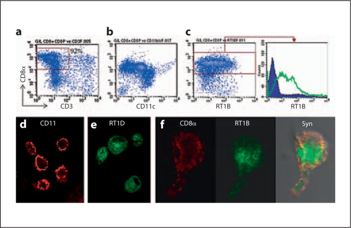
Background/aims: In an antiglomerular basement membrane glomerulonephritis (GN) model, GN-resistant Lewis (LEW) rats naturally recover from early glomerular inflammation (days 21-23). We have previously identified a glomeruli-infiltrating CD8α(+)CD11(high)MHC II(+) cell (GIL CD8α(+) cell) in GN-prone Wistar Kyoto (WKY) rats, which terminates glomerular inflammation through inducing T cell apoptosis prior to glomerular fibrosis at days 35-40. We investigated if GIL CD8α(+) cells were also associated with the recovery in LEW rats.

Methods: GIL CD8α(+) cells in LEW rats were characterized; their infiltration was observed in connection with T cell apoptosis in glomeruli.

Results: An influx of GIL CD8α(+) cells into inflamed glomeruli was confirmed in the immunized LEW rats at days 17-22, which was much earlier than days 28-35 in WKY rats. Notably, LEW rats had a GIL CD8α(+)CD11(high) subpopulation after day 17, while WKY rats lacked this population until after day 30. Analyses further revealed a large number of clustered apoptotic CD4(+) or CD3(+) T cells in the glomeruli during recovery (day 23) in LEW rats, as compared to day 35 (transition to fibrosis) in WKY rats. Thus, infiltration of GIL CD8α(+) cells coincided with decline of glomerular inflammation and T cell apoptosis during recovery in LEW rats. Isolated GIL CD8α(+) cells were able to infiltrate glomeruli in both WKY and LEW rats at day 20.

Conclusion: Our data revealed a strong association between GIL CD8a+ cells and recovery from early glomerular inflammation. It raises a possibility of involvement of GIL CD8a+ cells in the recovery.


Figures

Fig. 1
Fig. 1
Development of transient T cell-mediated anti-GBM GN in GN-resistant LEW rats in comparison to GN-prone WKY rats. a Time course of GN development after immunization with nephritogenic T cell epitope pCol(28–40). Each dot represents one individual. Severity of GN is expressed as glomerular injury score, from 0 (normal) to 4 (100% of glomeruli with fibrous crescentic lesions). Black dots = LEW; red dots = WKY; empty dots = WKY, fibrotic. Time frames for recovery in LEW (red letters) and transition to fibrosis in WKY rats (black letters) are indicated at the top. b Flow cytometry identification of glomeruli infiltrating CD4+ T cells. c Immunofluorescent micrographs show infiltrating CD4+ T cells (green) and ED-1+ macrophages (red) at days 15, 21, and 28 after immunization in GN-resistant LEW rats. Note that no CD4+ T cells and ED-1+ macrophages are present at day 28. Nuclei were counterstained by DAPI, and glomeruli outlined by arrowheads. ×300. For colors, see online version.
Fig. 2
Fig. 2
Identification of glomeruli-infiltrating CD8α+ cells (GIL CD8α+ cells) in GN-resistant LEW rats at day 21 after immunization with pCol(28–40). a Phase contrast micrograph shows various cells released from the glomeruli of LEW rats after digestion. b Flow cytometry reveals several morphologically different cell populations isolated from the glomeruli. Cells in gate R1 contain a GIL CD8α+ population. c–e Flow cytometry analyses based on R1 show a GIL CD8α+ population (red gate), which was CD8α+CD3 (c), CD11b/c+ (red gate in d) and RT1B+ (red gate in e). f Flow cytometry shows 42% of GIL CD8α+ cells were CD34+. g GIL CD8α+ cells did not express CD94 (upper panel) or TcRγδ (lower panel). For colors, see online version.
Fig. 3
Fig. 3
Characterization of GIL CD8α+ cells isolated from immunized LEW rats. a, b Flow cytometries show purified GIL CD8α+ cells to be CD3CD11c+. b Note two CD11c+ subpopulations: a majority of CD11high and a minority of CD11low. c The expression of various levels of MHC class II molecule (RT1B) in purified CD8α+ cells. The histogram on the right is based on the red gate in the left panel. Green line, antibody to RT1B (OX6), blue line, IgG control. d, e Confocal micrographs show surface CD11b/c (red), and surface/intracellular MHC II molecule RT1D (green) in CD8α+ cells. ×400. f Confocal micrograph demonstrates colocalization of RT1B (green) and CD8α in a GIL CD8α+ cell. ×600. Cells were cultured for one day before being processed for staining. For colors, see online version.
Fig. 4
Fig. 4
GIL CD8α+ cells of LEW rats are able to infiltrate inflamed glomeruli of both LEW and WKY rats at an early stage. a Fluorescence shows no CFSE+ cells in a glomerulus of a normal LEW rat. b Fluorescent micrographs show transferred CFSE+ GIL CD8α+ cells (green) in a glomerulus of immunized LEW rats. Glomeruli are outlined by arrowheads. c Immunofluorescence shows two transferred CFSE+CD8α+ cells (arrows) among other endogenous CD8α+ cells in an inflamed glomerulus of a LEW rat at day 20. ×300. d Summary of transferred CFSE+ cells in glomeruli in different groups. For colors, see online version.
Fig. 5
Fig. 5
CD8α+ cells infiltrate glomeruli earlier in GN-resistant LEW rats than in GN-prone WKY rats after immunization. a A group of confocal micrographs show GIL CD8α+ cells (red) in glomeruli after pCol(28–40) immunization as indicated. GBM (green) was counterstained with SR13. Average numbers of GIL CD8α+ cells per glomerulus are shown in figure 6a. Normal rats have 7.6 ± 5 cells/glomerulus. ×300. b Flow cytometry comparison of GIL CD8α+ cells between LEW and WKY after pCol(28–40) immunization as indicated; red gates indicate the population of CD8α+CD11high cells. Note that LEW rats had CD8α+CD11high population at day 20, which WKY rats lacked. See summarized data in figure 6b. For colors, see online version.
Fig. 6
Fig. 6
GIL CD8α+ cell infiltration is coincident with T cell apoptosis during GN recovery in LEW rats or transition to fibrosis in WKY rats. Summary of time courses for infiltration of GIL CD8α+ cells (a and b) and apoptosis in CD3+ or CD4+ cells (c and d) after immunization in GN-resistant LEW (red symbols) and GN-prone WKY rats (black symbols). Time frames for recovery stage in LEW rats and transition from inflammatory to fibrotic stage in WKY rats are indicated at the bottom. For colors, see online version.
Fig. 7
Fig. 7
Apoptosis in CD3+CD4+ cells in glomeruli of GN-resistant LEW during recovery stage. a Immunohistoperoxidase method detection of caspase-3-positive apoptotic cells in glomeruli of LEW and WKY as indicated. Clustered caspase 3+ cells are present in glomeruli of LEW at day 23, but not in WKY rats despite prominent glomerular inflammation in both strains. Clustered caspase 3+ cells are detectable in the periglomerular area of a glomerulus undergoing fibrosis in WKY at day 35. ×200. See summarized data for numbers of caspase+ cells in figure 6c. b Immunofluorescence shows colocalization of cytoplasmic caspase 3 (green) with cell surface CD3 (red) among clustered caspase 3+ cells in a glomerulus of a LEW rat at day 23. Arrows indicate the cells positive for both intracellular caspase 3 and surface CD3. Nuclei were counterstained with DAPI. ×400. c Flow cytometry analysis of isolated glomerular cells shows a significant size of apoptotic CD4+ T cell population (annexin V+ cells) in LEW but not in WKY rats at day 23. Gates R1 and R2 represent CD4+annexinV and CD4+annexinV+ populations. Histogram on the right shows comparison of expression level of annexin V between R1 and R2 populations. Percentages of annexin V+ cells among total CD4+ cells are summarized in figure 6d. For colors, see online version.

References

    1. Takasu N, Yamada T, Takasu M, Komiya I, Nagasawa Y, Asawa T, Shinoda T, Aizawa T, Koizumi Y. Disappearance of thyrotropin-blocking antibodies and spontaneous recovery from hypothyroidism in autoimmune thyroiditis. N Engl J Med. 1992;326:513–518. - PubMed
    1. Sultan Y, Rossi F, Kazatchkine MD. Recovery from anti-VIII:C (antihemophilic factor) autoimmune disease is dependent on generation of antiidiotypes against anti-VIII:C autoantibodies. Proc Natl Acad Sci USA. 1987;84:828–831. - PMC - PubMed
    1. McGeachy MJ, Stephens LA, Anderton SM. Natural recovery and protection from autoimmune encephalomyelitis: contribution of CD4+CD25+ regulatory cells within the central nervous system. J Immunol. 2005;175:3025–3032. - PubMed
    1. Tabi Z, McCombe AP, Pender MP. Antigen-specific down-regulation of myelin basic protein-reactive T cells during spontaneous recovery from experimental autoimmune encephalomyelitis: further evidence of apoptotic deletion of autoreactive T cells in the central nervous system. Int Immunol. 1995;7:967–973. - PubMed
    1. Arnold B, Schonrich G, Hammerling GJ. Multiple levels of peripheral tolerance. Immunol Today. 1993;14:12–14. - PubMed

Publication types