Clinical Trial
. 2012 Feb;20(2):443-55.
doi: 10.1038/mt.2011.237. Epub 2011 Nov 8.

Phase 1 gene therapy for Duchenne muscular dystrophy using a translational optimized AAV vector

Affiliations
Clinical Trial

Phase 1 gene therapy for Duchenne muscular dystrophy using a translational optimized AAV vector

Dawn E Bowles et al. Mol Ther. 2012 Feb.

Abstract

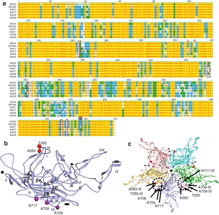
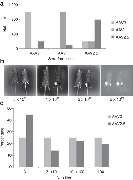
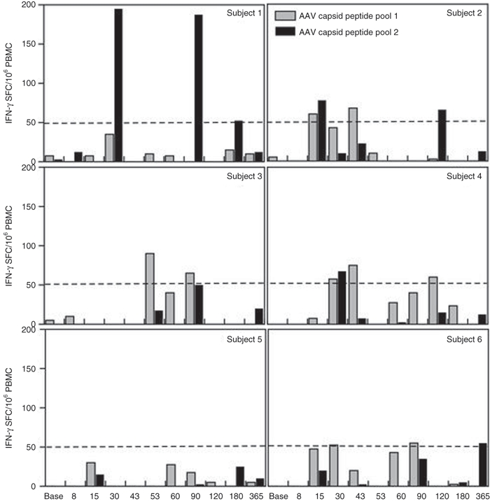
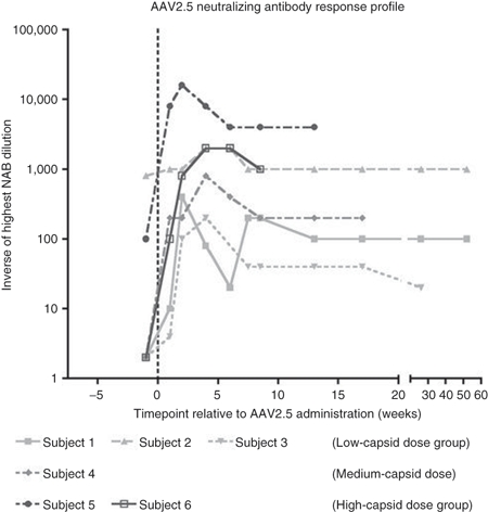
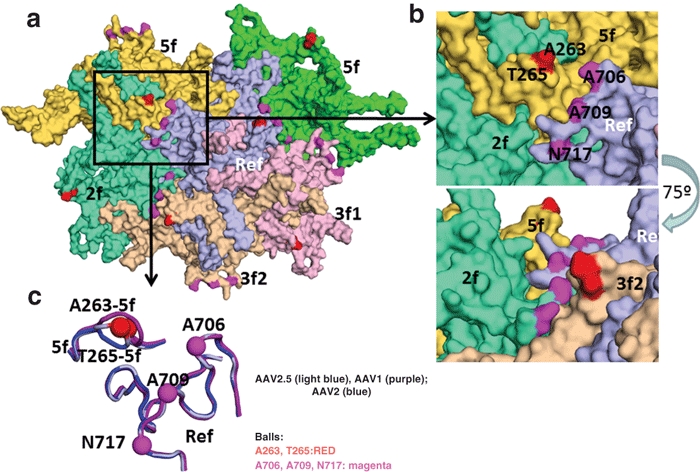
Efficient and widespread gene transfer is required for successful treatment of Duchenne muscular dystrophy (DMD). Here, we performed the first clinical trial using a chimeric adeno-associated virus (AAV) capsid variant (designated AAV2.5) derived from a rational design strategy. AAV2.5 was generated from the AAV2 capsid with five mutations from AAV1. The novel chimeric vector combines the improved muscle transduction capacity of AAV1 with reduced antigenic crossreactivity against both parental serotypes, while keeping the AAV2 receptor binding. In a randomized double-blind placebo-controlled phase I clinical study in DMD boys, AAV2.5 vector was injected into the bicep muscle in one arm, with saline control in the contralateral arm. A subset of patients received AAV empty capsid instead of saline in an effort to distinguish an immune response to vector versus minidystrophin transgene. Recombinant AAV genomes were detected in all patients with up to 2.56 vector copies per diploid genome. There was no cellular immune response to AAV2.5 capsid. This trial established that rationally designed AAV2.5 vector was safe and well tolerated, lays the foundation of customizing AAV vectors that best suit the clinical objective (e.g., limb infusion gene delivery) and should usher in the next generation of viral delivery systems for human gene transfer.


Figures

Figure 1
Figure 1
Amino acid candidates responsible for efficient skeletal muscle transduction. (a) Capsid amino acids of low skeletal muscle transducing serotypes (AAV2, AAV3) versus high skeletal muscle transducers (AAV1, AAV6, AAV7, AAV8, AAV9) were aligned using the Vector NTI program (Invitrogen). Alignments were examined for distinct amino acids of AAV2 from the others. See text for additional modeling criteria. Amino acids boxed or marked with arrows were deemed to be of interest. AAV2.5 is composed of the five amino acids indicated by * and @. (b) Location of the five amino acids on a single VP subunit which were modified in the AAV2.5 variant. Notice that the five amino acids are located on opposite positions of one subunit. (c) Location of the same five amino acids (circles and arrows) in the context of an assembled AAV capsid pentamer. Notice that the five amino acids are now in close proximity when two subunits are assembled. The five amino acid changes are located near the twofold axis of symmetry. AAV, adeno-associated virus.
Figure 2
Figure 2
Evaluation of skeletal muscle transduction of AAV2 mutants. (a) Skeletal muscle transduction of AAV1, AAV2, and AAV2.5 examined over time (days 3, 7, 21, 28, 42) using in vivo biophotonic imaging. The relative light units per region of interest in each injected mouse (n = 6) are graphed over time. (b) Graphical representation of quantity of emitted light from transduction of AAV2 and AAV2-Q325T/T329V. The relative light units per region of interest in each injected mouse leg (n = 6) are graphed over time. AAV, adeno-associated virus.
Figure 3
Figure 3
Neutralizing antibody analysis to AAV2.5. (a) Crossreactive Nab between AAV1, AAV2, and AAV2.5. C57 mice were immunized with 1 × 10¹⁰ particles of AAV/luc vectors via muscular injection. Thirty days later, sera from three mice was collected for Nab analysis. (b) The effect of AAV2 Nab on AAV2.5-induced transgene expression in vivo. Mice were immunized with AAV2/AAT viruses (left three panels) or not immunized (right panel), 2 months later, AAV2.5/luciferase (mouse right leg) and AAV2/luciferase (left leg) vectors with different dosages were applied in the same mice intramuscularly (5 × 10⁹ particles, 1 × 10¹⁰ particles, or 5 × 10¹⁰ particles), imaging was taken 6 weeks later post-luciferase vector injection. (c) Neutralizing antibody assay for human sera. The sera from 36 human subjects were detected for Nab against AAV2 and AAV2.5. AAV, adeno-associated virus.
Figure 4
Figure 4
Temporal T cell response to AAV2.5 capsid. Two capsid peptide pools were comprised of peptides spanning the AAV2.5 capsid sequence. Elispot assays measured interferon (IFN)-γ release upon peptide exposure, with the threshold for peripheral blood mononuclear cell (PBMC) recognition of an epitope within the peptide pool being 50 spots/106 PBMCs. Temporal responses are shown for subjects 001–006 in separate panels for the two peptide pools. AAV, adeno-associated virus.
Figure 5
Figure 5
Temporal neutralizing antibody response to AAV2.5. The temporal profile of the highest dilution of serum that neutralizes AAV2.5 transduction in vitro is shown for all six subjects. Subjects 1–3 received the same dose of AAV2.5 capsid, 6.6 × 10¹¹ capsid particles. Subject 4 received 3.3 × 10¹² capsid particles. Subjects 5 and 6 received 6.6 × 10¹² capsid particles due to administration of empty capsid placebo (see Table 2). AAV, adeno-associated virus.
Figure 6
Figure 6
Topology of AAV2.5 virion. Surface topology view of the five VP monomers (in different colors) immediately surrounding the reference monomer in light blue. The symmetry operation that brings the monomer into contact is given in the labels. The positions of the AAV2.5 residue mutations are colored as in Figure 1b,c. (b) Top panel is a close up of the boxed region in the a. The bottom panel shows the same image rotated by ~75° and shows that the region containing the 263/265 amino acids are raised on the capsid surface. (c) Image showing a surface view (same as in Figure 1) of the loop containing the 263/265 region, VR III and VR IX (close to the 706, 709, 717 mutations) for AAV1 (purple), AAV2 (blue), and AAV2.5 (light blue). The positions of the AAV2.5 mutants are shown in the balls and labeled. AAV, adeno-associated virus.

Comment in

References

    1. Koenig M, Hoffman EP, Bertelson CJ, Monaco AP, Feener C., and, Kunkel LM. Complete cloning of the Duchenne muscular dystrophy (DMD) cDNA and preliminary genomic organization of the DMD gene in normal and affected individuals. Cell. 1987;50:509–517. - PubMed
    1. Emery AE. The muscular dystrophies. Lancet. 2002;359:687–695. - PubMed
    1. Pichavant C, Aartsma-Rus A, Clemens PR, Davies KE, Dickson G, Takeda S.et al. (2011Current status of pharmaceutical and genetic therapeutic approaches to treat DMD Mol Ther 19830–840. - PMC - PubMed
    1. Gregorevic P, Allen JM, Minami E, Blankinship MJ, Haraguchi M, Meuse L.et al. (2006rAAV6-microdystrophin preserves muscle function and extends lifespan in severely dystrophic mice Nat Med 12787–789. - PMC - PubMed
    1. Wang B, Li J, Fu FH., and, Xiao X. Systemic human minidystrophin gene transfer improves functions and life span of dystrophin and dystrophin/utrophin-deficient mice. J Orthop Res. 2009;27:421–426. - PubMed

Publication types

MeSH terms