. 2012 Feb 15;21(4):776-83.
doi: 10.1093/hmg/ddr509. Epub 2011 Nov 7.

Next generation sequencing identifies mutations in Atonal homolog 7 (ATOH7) in families with global eye developmental defects

Affiliations

Next generation sequencing identifies mutations in Atonal homolog 7 (ATOH7) in families with global eye developmental defects

Kamron Khan et al. Hum Mol Genet. .

Abstract

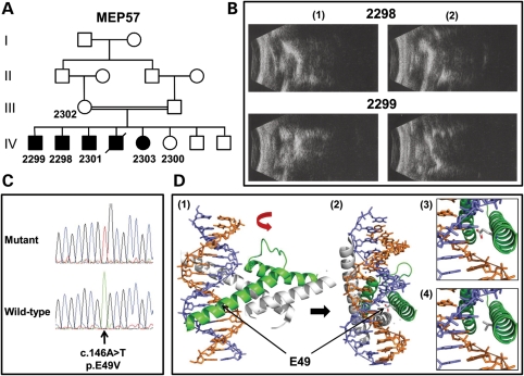
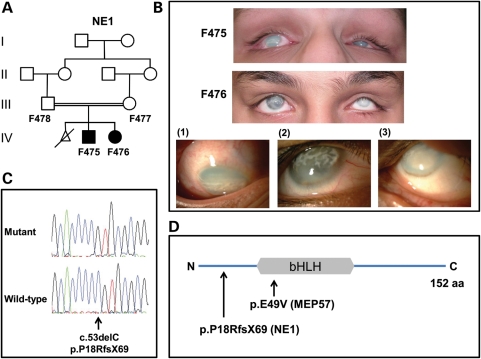
The atonal homolog 7 (ATOH7) gene encodes a transcription factor involved in determining the fate of retinal progenitor cells and is particularly required for optic nerve and ganglion cell development. Using a combination of autozygosity mapping and next generation sequencing, we have identified homozygous mutations in this gene, p.E49V and p.P18RfsX69, in two consanguineous families diagnosed with multiple ocular developmental defects, including severe vitreoretinal dysplasia, optic nerve hypoplasia, persistent fetal vasculature, microphthalmia, congenital cataracts, microcornea, corneal opacity and nystagmus. Most of these clinical features overlap with defects in the Norrin/β-catenin signalling pathway that is characterized by dysgenesis of the retinal and hyaloid vasculature. Our findings document Mendelian mutations within ATOH7 and imply a role for this molecule in the development of structures at the front as well as the back of the eye. This work also provides further insights into the function of ATOH7, especially its importance in retinal vascular development and hyaloid regression.


Figures

Figure 1.
Figure 1.
Analysis of MEP57. (A) Pedigree structure for family MEP57. The individuals from whom DNA is available are numbered. (B) B-scan ultrasound examination of the right (1) and left (2) eyes from patients 2298 (aged 33 years) and 2299 (aged 24 years). Note a reduced axial length (microphthalmia) together with disorganization of the vitreous and chronic retinal detachments. (C) Sequence chromatograms highlighting the ATOH7 mutation in an affected patient from MEP57 and the corresponding wild-type sequence from a normal individual. (D) Molecular modelling of the bHLH domain of human ATOH7 wild-type and p.E49V. Ribbon representation of the bHLH domain in ATOH7 (in green) dimerized with the transcription factor E2-alpha (in grey) and bound to double-stranded DNA (purple and orange helix) as a lateral (1) and, following a 90° rotation, frontal (2) view. Note residue E49 in the wild-type sequence is in close connection with the DNA helix. The proximity of the E49 (3) and V49 (4) residues to the DNA helix shows that the V49 is more distant and so is less likely to interact with the DNA.
Figure 2.
Figure 2.
Analysis of NE1. (A) Pedigree structure for family NE1. The individuals from whom DNA is available are numbered. (B) Current photos of the anterior segment of affected family members F475 (aged 16 years) and F476 (aged 14 years). Detailed views of the right eye of patients F475 (1) and F476 (2) as well as the left eye of patient F476 (3) are shown. Note reduced globe size (microphthalmia), diffuse corneal haze with additional superficial, predominantly interpalpebral, discrete opacity secondary to calcium deposition (band keratopathy). The observed ocular deviation (strabismus) is a result of the poor vision. There are no signs associated with significant anterior segment inflammation. (C) Sequence chromatograms highlighting the ATOH7 mutation in an affected patient from NE1 and the corresponding wild-type sequence from a normal individual. (D) Diagrammatic representation of ATOH7 (NP_660161). The position of the bHLH domain and the mutations relative to the full-length 152 amino acid protein are shown.

References

    1. Cepko C.L., Austin C.P., Yang X., Alexiades M., Ezzeddine D. Cell fate determination in the vertebrate retina. Proc. Natl Acad. Sci. USA. 1996;93:589–595. - PMC - PubMed
    1. Tomita K., Moriyoshi K., Nakanishi S., Guillemot F., Kageyama R. Mammalian achaete-scute and atonal homologs regulate neuronal versus glial fate determination in the central nervous system. EMBO J. 2000;19:5460–5472. - PMC - PubMed
    1. Brown N.L., Dagenais S.L., Chen C.M., Glaser T. Molecular characterization and mapping of ATOH7, a human atonal homolog with a predicted role in retinal ganglion cell development. Mamm. Genome. 2002;13:95–101. - PMC - PubMed
    1. Murre C., McCaw P.S., Vaessin H., Caudy M., Jan L.Y., Jan Y.N., Cabrera C.V., Buskin J.N., Hauschka S.D., Lassar A.B., et al. Interactions between heterologous helix-loop-helix proteins generate complexes that bind specifically to a common DNA sequence. Cell. 1989;58:537–544. - PubMed
    1. Ferre-D'Amare A.R., Prendergast G.C., Ziff E.B., Burley S.K. Recognition by Max of its cognate DNA through a dimeric b/HLH/Z domain. Nature. 1993;363:38–45. - PubMed

Publication types

Associated data