Clinical Trial
. 2011 Dec;52(12):1878-85.
doi: 10.2967/jnumed.111.095596. Epub 2011 Nov 8.

124I-huA33 antibody uptake is driven by A33 antigen concentration in tissues from colorectal cancer patients imaged by immuno-PET

Affiliations
Clinical Trial

124I-huA33 antibody uptake is driven by A33 antigen concentration in tissues from colorectal cancer patients imaged by immuno-PET

Joseph A O'Donoghue et al. J Nucl Med. 2011 Dec.

Abstract

The primary aim of this analysis was to examine the quantitative features of antibody-antigen interactions in tumors and normal tissue after parenteral administration of antitumor antibodies to human patients.

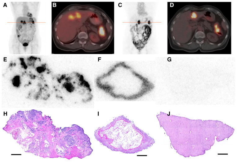
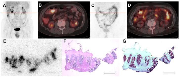
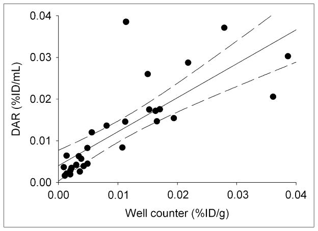
Methods: Humanized anti-A33 antibody (10 mg) labeled with the positron-emitting radionuclide (124)I ((124)I-huA33) was injected intravenously in 15 patients with colorectal cancer. Clinical PET/CT was performed approximately 1 wk later, followed by a detailed assay of surgically removed tissue specimens including radioactivity counting, autoradiography, immunohistochemistry, and antigen density determination.

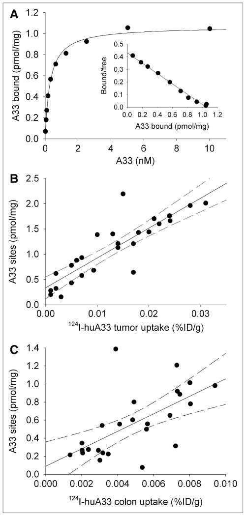
Results: PET/CT showed high levels of antibody targeting in tumors and normal bowel. In tissue specimens, the spatial distribution of (124)I-huA33 conformed to that of A33 antigen, and there was a linear relationship between the amount of bound antibody and antigen concentration. Antibody uptake was high in 1- to 2-mm regions of antigen-positive tumor cells (mean, ~0.05 percentage injected dose per gram) and in antigen-positive normal colonic mucosa (mean, ~0.03 percentage injected dose per gram). The estimated binding site occupancy for tumor and normal colon was 20%-50%.

Conclusion: The in vivo biodistribution of (124)I-huA33 in human patients 1 wk after antibody administration was determined by A33 antigen expression. Our data imply that the optimal strategy for A33-based radioimmunotherapy of colon cancer will consist of a multistep treatment using a radionuclide with short-range (α- or β-particle) emissions.


Conflict of interest statement

No other potential conflict of interest relevant to this article was reported.

Figures

FIGURE 1
FIGURE 1
(A–B) 18F-FDG PET/CT scan acquired immediately before administration of 124I-huA33: coronal maximum-intensity-projection image (A) and transaxial PET/CT slice (B) through line of A. Images show pathologic uptake in primary (splenic flexure of colon) and metastatic (liver) lesions together with normal physiologic accumulation. (C–D) Clinical PET/CT scan acquired 162 h after administration of 395 MBq (10.7 mCi) of 124I-huA33: coronal maximum-intensity-projection (C) and transaxial PET/CT slice (D) through line of C. Intense focal uptake is visible in primary and metastatic tumor together with significant uptake throughout normal intestine. (E–J) DAR (E–G) and hematoxylin and eosin–stained (H–J) tissue sections corresponding to features observed in clinical image: primary tumor (E and H), normal colon (F and I), and normal liver (G and J). In tumor section, 124I-huA33 is localized primarily in regions of viable tumor cells with little stromal uptake. Normal colonic mucosa has high and uniform uptake. Little to no uptake is present in normal liver. Scale bars = 2 mm.
FIGURE 2
FIGURE 2
(A–B) 18F-FDG PET/CT scan acquired immediately before administration of 124I-huA33 shows pathologic uptake in lesion in transverse colon near hepatic flexure together with normal physiologic accumulation. (C–D) PET/CT scan acquired 182 h after administration of 140 MBq (3.8 mCi) of 124I-huA33 shows focal uptake in tumor (compare B and D) and throughout normal intestine. (E–G) Contiguously aligned sections of tumor visualized in D assessed by 124I-DAR (E), hematoxylin and eosin staining (F), and immunohistochemical staining (G) for A33 antigen. 124I-huA33 localized only in small (~1 mm in dimension) regions of viable antigen-positive tumor and not in antigen-negative stroma. Scale bars = 2 mm.
FIGURE 3
FIGURE 3
(A) Example of A33 saturation binding curve for membranes derived from surgically removed tumor sample together with Scatchard transformed data plot (insert). Mean Kd ± SE, 0.31 ± 0.02 nM; mean Bmax ± SE, 1.07 ± 0.02 pmol/mg. (B) Scatterplot of A33 antigen density for membranes derived from tumor homogenates vs. 124I-huA33 uptake as determined by well counter measurements. Predicted 95% confidence limits are shown as dashed lines. Regression equation, y = 59 (SE, 7)x + 0.3 (SE, 0.1) (n = 26; r2 = 0.75). (C) Scatterplot of A33 antigen density for membranes derived from normal colon homogenates vs. 124I-huA33 uptake determined by well counter measurements. Regression equation, y = 97 (SE, 25)x + 0.09 (SE, 0.1) (n = 25; r2 = 0.40).
FIGURE 4
FIGURE 4
Scatterplot of tissue %ID/mL estimated using DAR vs. well counter measurements (%ID/g) of intact tissue samples. Regression equation, y = 0.81 (SE, 0.12)x + 0.004 (SE, 0.002) (n = 29; r2 = 0.62).

References

    1. Garinchesa P, Sakamoto J, Welt S, Real FX, Rettig WJ, Old LJ. Organ-specific expression of the colon cancer antigen A33, a cell surface target for antibody-based therapy. Int J Oncol. 1996;9:465–471. - PubMed
    1. Johnstone CN, White SJ, Tebbutt NC, et al. Analysis of the regulation of the A33 antigen gene reveals intestine-specific mechanisms of gene expression. J Biol Chem. 2002;277:34531–34539. - PubMed
    1. Sakamoto J, Kojima H, Kato J, Hamashima H, Suzuki H. Organ-specific expression of the intestinal epithelium-related antigen A33, a cell surface target for antibody-based imaging and treatment in gastrointestinal cancer. Cancer Chemother Pharmacol. 2000;46:S27–S32. - PubMed
    1. Heath JK, White SJ, Johnstone CN, et al. The human A33 antigen is a transmembrane glycoprotein and a novel member of the immunoglobulin superfamily. Proc Natl Acad Sci USA. 1997;94:469–474. - PMC - PubMed
    1. Ackerman ME, Chalouni C, Schmidt MM, et al. A33 antigen displays persistent surface expression. Cancer Immunol Immunother. 2008;57:1017–1027. - PMC - PubMed

Publication types

MeSH terms

Substances