. 2011 Dec 30;286(52):44606-19.
doi: 10.1074/jbc.M111.278903. Epub 2011 Nov 8.

O-GlcNAcylation, novel post-translational modification linking myocardial metabolism and cardiomyocyte circadian clock

Affiliations

O-GlcNAcylation, novel post-translational modification linking myocardial metabolism and cardiomyocyte circadian clock

David J Durgan et al. J Biol Chem. .

Abstract

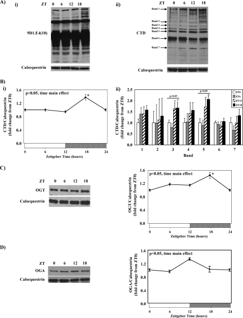
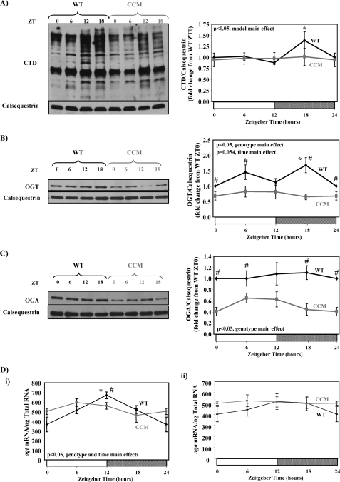
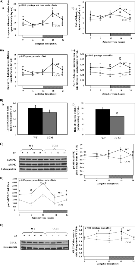
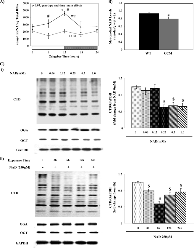
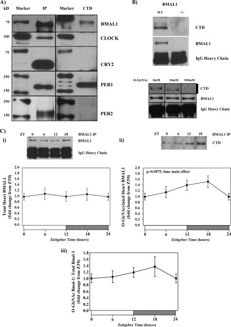
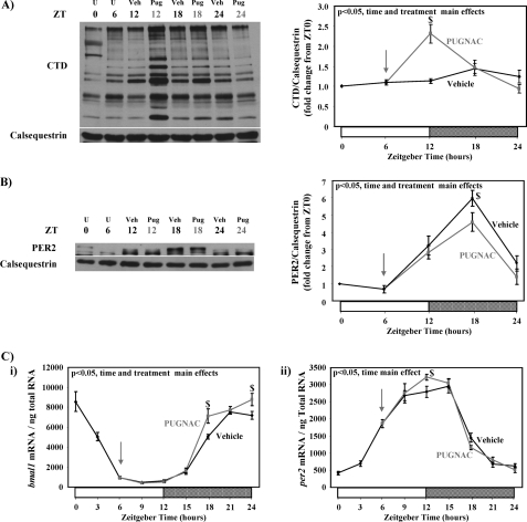
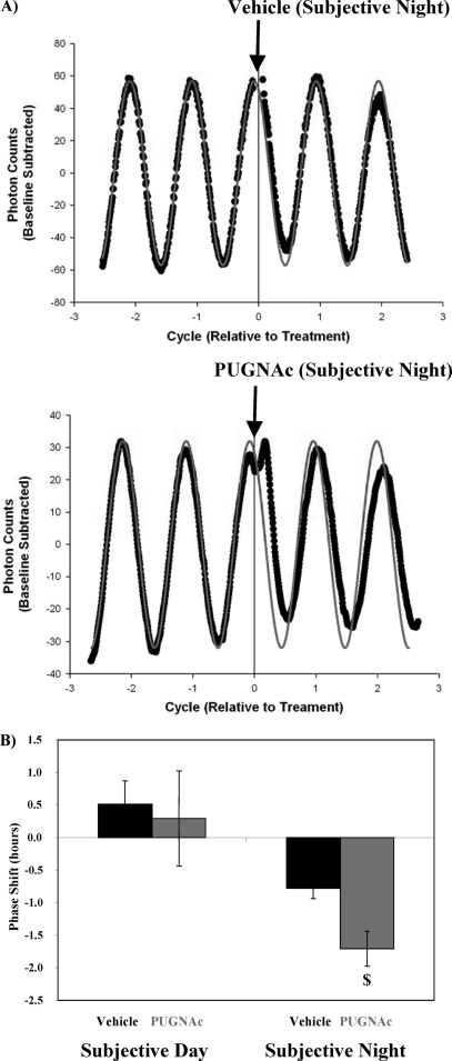
The cardiomyocyte circadian clock directly regulates multiple myocardial functions in a time-of-day-dependent manner, including gene expression, metabolism, contractility, and ischemic tolerance. These same biological processes are also directly influenced by modification of proteins by monosaccharides of O-linked β-N-acetylglucosamine (O-GlcNAc). Because the circadian clock and protein O-GlcNAcylation have common regulatory roles in the heart, we hypothesized that a relationship exists between the two. We report that total cardiac protein O-GlcNAc levels exhibit a diurnal variation in mouse hearts, peaking during the active/awake phase. Genetic ablation of the circadian clock specifically in cardiomyocytes in vivo abolishes diurnal variations in cardiac O-GlcNAc levels. These time-of-day-dependent variations appear to be mediated by clock-dependent regulation of O-GlcNAc transferase and O-GlcNAcase protein levels, glucose metabolism/uptake, and glutamine synthesis in an NAD-independent manner. We also identify the clock component Bmal1 as an O-GlcNAc-modified protein. Increasing protein O-GlcNAcylation (through pharmacological inhibition of O-GlcNAcase) results in diminished Per2 protein levels, time-of-day-dependent induction of bmal1 gene expression, and phase advances in the suprachiasmatic nucleus clock. Collectively, these data suggest that the cardiomyocyte circadian clock increases protein O-GlcNAcylation in the heart during the active/awake phase through coordinated regulation of the hexosamine biosynthetic pathway and that protein O-GlcNAcylation in turn influences the timing of the circadian clock.


Figures

FIGURE 1.
FIGURE 1.
Diurnal variations in mouse heart protein O-GlcNAcylation using the anti-O-GlcNAc antibody 9D1.E4(10) (i) or CTD (ii) (A) is shown. Densitometric analysis of total protein O-GlcNAcylation (i) as well as seven distinct O-GlcNAc protein bands (ii) in the mouse heart (B) is shown. Diurnal variations in OGT (C) and OGA (D) protein levels in the mouse heart are shown. Hearts were isolated at the dark-to-light phase transition (ZT 0), middle of the light phase (ZT 6), light-to-dark phase transition (ZT 12), and middle of the dark phase (ZT 18); ZT 0 and ZT 24 are identical data points. ZT represents zeitgeber time. Data are shown as the mean ± S.E. for between 14 and 20 separate hearts within each group. The main effects for time are indicated in the top left hand corner of the figure panels. *, p < 0.05 for a specific time point versus the trough (i.e. lowest) value.
FIGURE 2.
FIGURE 2.
Diurnal variations in protein O-GlcNAcylation (A), OGT (B), and OGA (C) protein levels as well as ogt and oga mRNA levels (D) in WT versus CCM hearts. Hearts were isolated at the dark-to-light phase transition (ZT 0), middle of the light phase (ZT 6), light-to-dark phase transition (ZT 12), and middle of the dark phase (ZT 18); ZT 0 and ZT 24 are identical data points. ZT represents zeitgeber time. Data are shown as the mean ± S.E. for between 11 and 20 separate hearts within each group. Main effects for model, time, or genotype are indicated in the top or bottom left-hand corner of the figure panels. *, p < 0.05 for a specific time point versus the trough (i.e. lowest) value. #, p < 0.05 for WT versus CCM at a distinct zeitgeber time.
FIGURE 3.
FIGURE 3.
Time-of-day- and cardiomyocyte circadian clock-dependent differences in parameters influencing entry of carbon into the HBP. Diurnal variations in rates of glucose oxidation (i), glycolysis (ii and iii), and glycogen synthesis (iv) in ex vivo perfused in WT versus CCM hearts are shown (A). Rates of lactate oxidation (i) and glucose uptake (ii) for WT versus CCM hearts at ZT18 are shown (B). Diurnal variations in P-AMPK levels in WT versus CCM hearts are shown (C). Diurnal variations in glul mRNA levels in WT versus CCM hearts (D) are shown. Diurnal variations in GLUL protein levels in WT versus CCM hearts (E) are shown. Hearts were isolated at the dark-to-light phase transition (ZT 0), middle of the light phase (ZT6), light-to-dark phase transition (ZT 12), and middle of the dark phase (ZT 18); ZT 0 and ZT 24 are identical data points. ZT represents zeitgeber time. Data are shown as the mean ± S.E. for between 7 and 15 separate hearts within each group. Main effects for model, time, or genotype are indicated in the top left-hand corner of the figure panels. *, p < 0.05 for a specific time point versus the trough (i.e. lowest) value. #, p < 0.05 for WT versus CCM at a distinct ZT.
FIGURE 4.
FIGURE 4.
Influence of NAD on protein O-GlcNAcylation. Diurnal variations in nampt mRNA levels in WT versus CCM hearts (A) are shown. NAD levels in WT versus CCM hearts at ZT18 (B) are shown. Concentration- and time-dependent effects of NAD on protein O-GlcNAcylation as well as OGT and OGA expression in neonatal rat ventricular cardiomyocytes (C) are shown. Hearts were isolated at the dark-to-light phase transition (ZT 0), middle of the light phase (ZT 6), light-to-dark phase transition (ZT 12), and middle of the dark phase (ZT 18); ZT 0 and ZT 24 are identical data points. ZT represents zeitgeber time. Data are shown as the mean ± S.E., for between five and six separate hearts/experiments within each group. Main effects for model, time, or genotype are indicated in the top left-hand corners of figure panels. *, p < 0.05 for a specific time point versus the trough (i.e. lowest) value. #, p < 0.05 for WT versus CCM at a distinct ZT. $, p < 0.05 for a specific NAD challenge compared with control.
FIGURE 5.
FIGURE 5.
Identification of O-GlcNAc-modified clock components. Screening clock components (BMAL1, CLOCK, CRY2, PER1, and PER2) for O-GlcNAc modification in mouse livers is shown (A). The absence of BMAL1 O-GlcNAc modification in BMAL1 null mouse livers or after CTD-binding blockage with O-GlcNAc (10 or 100 mm) (B) is shown. Diurnal variations in Bmal1 O-GlcNAcyation in hearts (D) is shown. Hearts were isolated at the dark-to-light phase transition (ZT 0), middle of the light phase (ZT 6), light-to-dark phase transition (ZT 12), and middle of the dark phase (ZT 18); ZT 0 and ZT 24 are identical data points. ZT represents zeitgeber time. Data are shown as the mean ± S.E. for between 6 and 14 separate hearts/livers within each group. Main effects for time are indicated in the top left-hand corners of figure panels. *, p < 0.05 for a specific time point versus the trough (i.e. lowest) value. IP, immunoprecipitation.
FIGURE 6.
FIGURE 6.
Influence of PUGNAc administration on levels of total protein O-GlcNAcylation (A) and protein expression of PER2 (B) as well as bmal1 and per2 gene expression (C) in mouse hearts. Mice were treated with PUGNAc (20 mg/kg intraperitoneally) at ZT 6 (indicated by an arrow) after which hearts were isolated at ZT 12, ZT 18, and ZT 24. Vehicle-treated mice served as controls. Hearts were isolated at the dark-to-light phase transition (ZT 0), middle of the light phase (ZT 6), light-to-dark phase transition (ZT 12), and middle of the dark phase (ZT 18); ZT 0 and ZT 24 are identical data points. The same samples were utilized for both protein and mRNA measurements. ZT represents zeitgeber time; U represents untreated (i.e. before vehicle or PUGNAc treatment); Veh represents vehicle treatment; Pug represents PUGNAc treatment. Data are shown as the mean ± S.E. for between five and six separate hearts within each group. Main effects for model, time, or treatment are indicated in the top left-hand corners of figure panels. *, p < 0.05 for a specific time point versus the trough (i.e. lowest) value. $, p < 0.05 for a PUGNAc treated versus vehicle at a distinct zeitgeber time.
FIGURE 7.
FIGURE 7.
Influence of OGA inhibition on circadian rhythms in luciferase for SCN isolated from Per2::Luc transgenic mice; representative raw data (gray line indicates predicted oscillation, black circles represent data obtained; A) and calculated phase shifts (B). SCN cultures were treated with vehicle (water) or PUGNAc (50 μm) either during the subjective day or subjective night, after which the effects on the phase of luminescence oscillations were determined (i.e. phase shift). PUGNAc challenge during the subjective night resulted in phase advances, whereas PUGNAc challenge during the subjective day had no effects on phase. Data are shown as the mean ± S.E. for between three and eight separate SCNs within each group. $, p < 0.05 for a PUGNAc treated versus vehicle at a distinct time in the circadian cycle.
FIGURE 8.
FIGURE 8.
Hypothetical model for the interaction between myocardial metabolism, protein O-GlcNAcylation, and the cardiomyocyte circadian clock. Genes/proteins within black boxes are considered cardiomyocyte circadian clock regulated.

References

    1. Durgan D. J., Young M. E. (2010) Circ. Res. 106, 647–658 - PMC - PubMed
    1. Bray M. S., Young M. E. (2009) Obes. Rev. 10, 6–13 - PubMed
    1. Turek F. W., Joshu C., Kohsaka A., Lin E., Ivanova G., McDearmon E., Laposky A., Losee-Olson S., Easton A., Jensen D. R., Eckel R. H., Takahashi J. S., Bass J. (2005) Science 308, 1043–1045 - PMC - PubMed
    1. Shimba S., Ishii N., Ohta Y., Ohno T., Watabe Y., Hayashi M., Wada T., Aoyagi T., Tezuka M. (2005) Proc. Natl. Acad. Sci. U.S.A. 102, 12071–12076 - PMC - PubMed
    1. Young M. E., Wilson C. R., Razeghi P., Guthrie P. H., Taegtmeyer H. (2002) J. Mol. Cell. Cardiol. 34, 223–231 - PubMed

Publication types

MeSH terms