. 2011;6(11):e26794.
doi: 10.1371/journal.pone.0026794. Epub 2011 Nov 1.

The molecular basis for the broad substrate specificity of human sulfotransferase 1A1

Affiliations

The molecular basis for the broad substrate specificity of human sulfotransferase 1A1

Ilana Berger et al. PLoS One. 2011.

Abstract

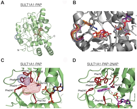
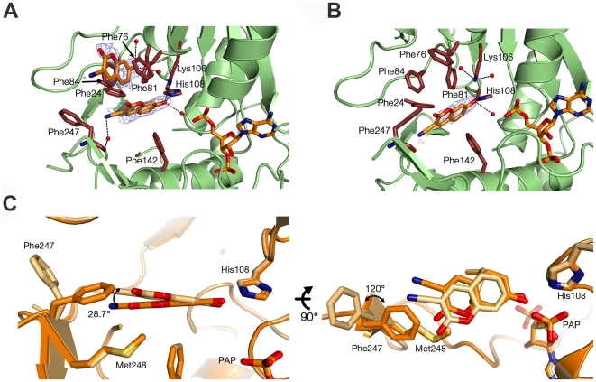
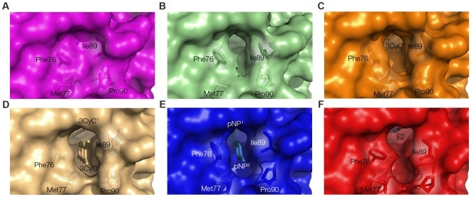
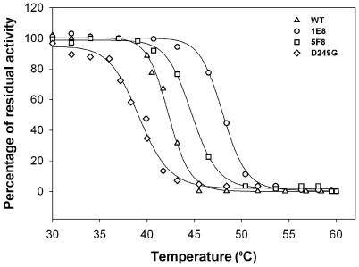
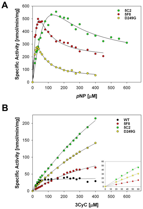
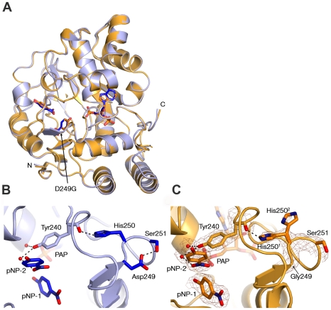
Cytosolic sulfotransferases (SULTs) are mammalian enzymes that detoxify a wide variety of chemicals through the addition of a sulfate group. Despite extensive research, the molecular basis for the broad specificity of SULTs is still not understood. Here, structural, protein engineering and kinetic approaches were employed to obtain deep understanding of the molecular basis for the broad specificity, catalytic activity and substrate inhibition of SULT1A1. We have determined five new structures of SULT1A1 in complex with different acceptors, and utilized a directed evolution approach to generate SULT1A1 mutants with enhanced thermostability and increased catalytic activity. We found that active site plasticity enables binding of different acceptors and identified dramatic structural changes in the SULT1A1 active site leading to the binding of a second acceptor molecule in a conserved yet non-productive manner. Our combined approach highlights the dominant role of SULT1A1 structural flexibility in controlling the specificity and activity of this enzyme.


Conflict of interest statement

Competing Interests: The authors have declared that no competing interests exist.

Figures

Figure 1
Figure 1. The overall structure of SULT1A1-PAP and flexibility of the SULT1A1 binding pocket.
(A) View of SULT1A1-PAP with the PAP molecule colored in brown and the gating loop (residues 86–90) colored in blue. (B) Structural flexibility of SULT1A1 is demonstrated by the overlapping of the donor and acceptor binding pockets of all newly and previously determined SULT1A1 structures (see text for details). The colors of the gating loop, the donor and the acceptors are as follows: SULT1A1-PAP-2NAP-pink, SULT1A1-PAP-green, SULT1A1-PAP-3CyC2-orange, SULT1A1-PAP-pNP-blue (PDB code 1LS6), SULT1A1-PAP-E2-red (PDB code 2D06) (C) SULT1A1 in complex with PAP shows the formation of a SULT1A1 empty acceptor binding site comprising pocket-1 and pocket-2, depicted as red and blue mesh blobs, respectively. (D) The structure of SULT1A1-PAP-2NAP. The 2NAP acceptor and PAP donor are colored in pink and the 2NAP is outlined with a FO-FC electron omit map contoured at 2.5ó. Key residues in panels C and D are colored red.
Figure 2
Figure 2. The molecular basis of SULT1A1 3CyC substrate inhibition.
Structure of SULT1A1 in complex with PAP and two molecules of 3CyC (SULT1A1-PAP-3CyC2, A) or one molecule of 3CyC (SULT1A1-PAP-3CyC1, B) bound at the acceptor-binding site. Key residues are highlighted in red and the 3CyC molecules are outlined with FO-FC electron omit maps contoured at 2.5ó. (C) Superposition of 3CyC molecules and key residues within the SULT1A1-PAP-3CyC1 and SULT1A1-PAP-3CyC2 structures indicating dramatic movements of the 3CyC cyano group and of Phe247.
Figure 3
Figure 3. Surface representation of the binding pocket and pore size of the different SULT1A1 structures.
Ligands and binding pockets are colored according to the displayed SULT1A1 structure: SULT1A1 in complex with PAP and 2NAP (A, pink), PAP (B, green), PAP and 3CyC1 (C, containing one 3CyC molecule, orange), PAP and 3CyC2 (D, containing two 3CyC molecules, grey), PAP and pNP (E, blue) or PAP and E2 (F, red). Key residues are highlighted in stick-form. Newly determined structures are A–D.
Figure 4
Figure 4. Heat inactivation curves for the WT SULT1A1 protein and the 5C2, 5F8 and D249G SULT1A1 mutants.
The 1E8 mutant was isolated after the first round of evolution for increased thermostability and the 5F8 was isolated after the second round of evolution for increased catalytic activity. The decrease in stability of the 5F8 mutant, relative to 1E8, and the D249G mutant, relative to the WT protein, highlight the trade-off between thermostability and the acquisition of new catalytic properties (see text for details).
Figure 5
Figure 5. Kinetic analysis of the sulfate transfer activity of the WT SULT1A1 protein and the 5C2, 5F8 and D249G SULT1A1 mutants to pNP (A) and 3CyC (B).
Each data point represents the mean of three independent experiments. The lines in A represent fit to data obtained with pNP of equation 1 (see Material and Methods) adopted from ref. 21, taking into account the inhibition seen at high pNP concentrations. The lines in B represent fit to Michaelis-Menten equation of data obtained with 3CyC or linear fit to data obtained at low 3CyC concentrations (inset). The kcat/KM parameters derived from the fits are 1208, 466, 149 and 308 for the WT, 5C2, 5F8 and D249G, respectively, presented as sec−1 M−1.
Figure 6
Figure 6. Comparison of the SULT1A1 and SULT1A1-D249G structures in complex with PAP and pNP.
(A) Structural superposition of SULT1A1 (PDB code 1LS6, light blue) and SULT1A1-D249G mutant (gold). (B–C) View of SULT1A1 (B) and the SULT1A1-D249G mutant (C) ligand pocket and the stabilizing loop containing Asp249 or Gly249 (rotamers are indicated with superscript). SULT1A1-D249G key residues and pNP molecule are outlined with a 2FO-FC electron density map contoured at 1.6ó.

References

    1. Chapman E, Best MD, Hanson SR, Wong CH. Sulfotransferases: structure, mechanism, biological activity, inhibition, and synthetic utility. Angew Chem Int Ed Engl. 2004;43:3526–3548. - PubMed
    1. Gamage N, Barnett A, Hempel N, Duggleby RG, Windmill KF, et al. Human sulfotransferases and their role in chemical metabolism. Toxicol Sci. 2006;90:5–22. - PubMed
    1. Negishi M, Pedersen LG, Petrotchenko E, Shevtsov S, Gorokhov A, et al. Structure and function of sulfotransferases. Arch Biochem Biophys. 2001;390:149–157. - PubMed
    1. Wang Y, Spitz MR, Tsou AM, Zhang K, Makan N, et al. Sulfotransferase (SULT) 1A1 polymorphism as a predisposition factor for lung cancer: a case-control analysis. Lung Cancer. 2002;35:137–142. - PubMed
    1. Blanchard RL, Freimuth RR, Buck J, Weinshilboum RM, Coughtrie MW. A proposed nomenclature system for the cytosolic sulfotransferase (SULT) superfamily. Pharmacogenetics. 2004;14:199–211. - PubMed

Publication types

LinkOut - more resources