. 2011;6(11):e27035.
doi: 10.1371/journal.pone.0027035. Epub 2011 Nov 1.

Bacillus subtilis MreB orthologs self-organize into filamentous structures underneath the cell membrane in a heterologous cell system

Affiliations

Bacillus subtilis MreB orthologs self-organize into filamentous structures underneath the cell membrane in a heterologous cell system

Felix Dempwolff et al. PLoS One. 2011.

Abstract

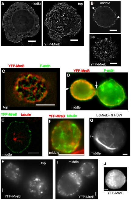
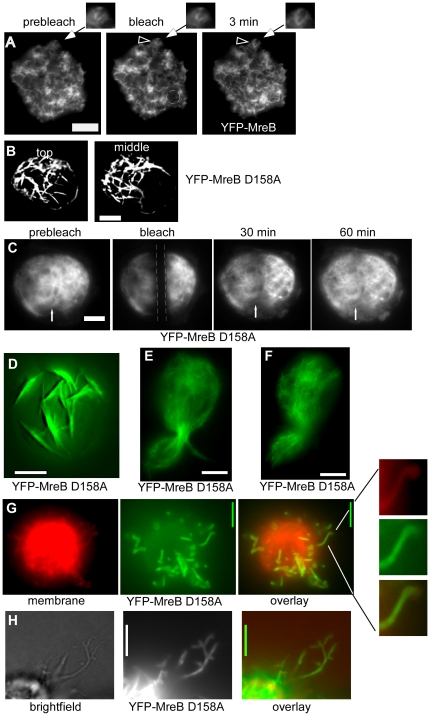
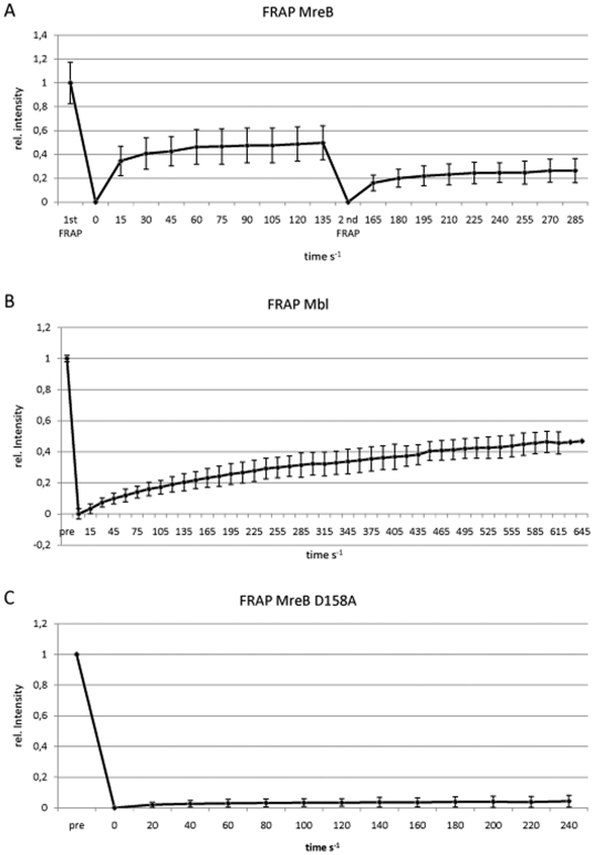
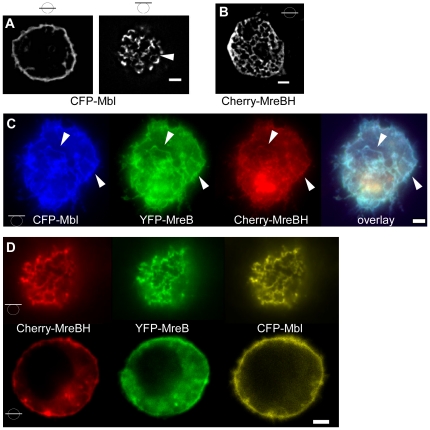
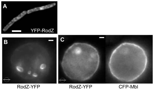
Actin-like bacterial cytoskeletal element MreB has been shown to be essential for the maintenance of rod cell shape in many bacteria. MreB forms rapidly remodelling helical filaments underneath the cell membrane in Bacillus subtilis and in other bacterial cells, and co-localizes with its two paralogs, Mbl and MreBH. We show that MreB localizes as dynamic bundles of filaments underneath the cell membrane in Drosophila S2 Schneider cells, which become highly stable when the ATPase motif in MreB is modified. In agreement with ATP-dependent filament formation, the depletion of ATP in the cells lead to rapid dissociation of MreB filaments. Extended induction of MreB resulted in the formation of membrane protrusions, showing that like actin, MreB can exert force against the cell membrane. Mbl also formed membrane associated filaments, while MreBH formed filaments within the cytosol. When co-expressed, MreB, Mbl and MreBH built up mixed filaments underneath the cell membrane. Membrane protein RodZ localized to endosomes in S2 cells, but localized to the cell membrane when co-expressed with Mbl, showing that bacterial MreB/Mbl structures can recruit a protein to the cell membrane. Thus, MreB paralogs form a self-organizing and dynamic filamentous scaffold underneath the membrane that is able to recruit other proteins to the cell surface.


Conflict of interest statement

Competing Interests: The authors have declared that no competing interests exist.

Figures

Figure 1
Figure 1. Expression of YFP-MreB or mutant versions in S2 cells.
A) Wild type YFP-MreB filaments, shown are a middle plane and top plane of a Z-stack. Triangles indicate bundles of filaments from which a single filament (or thin bundle of filaments) emanates. B) Middle and top planes of a 3D deconvoluted Z-stack of a cell expressing wild type YFP-MreB. C–D) Immunofluorescence of cell expressing YFP-MreB, using phalloidin as stain for actin filaments. Triangles indicate positions of actin filaments that lack any detectable YFP-MreB fluorescence. E–F) Immunofluorescence of cell expressing YFP-MreB, using anti Drosophila tubulin antiserum to stain for tubulin filaments. G) E. coli MreB (with an internal RFP) expressed in S2 cells, shown is the middle plane. Triangle indicates MreB filaments extending from the end of a filament bundle. H–I) Cells were depleted for ATP by the addition of FCCP, H–I) 20 min after addition, J) 90 min after addition (middle plane is shown). White bars 2 µm (A,B, G) or 5 µm (C–F) respectively, grey bars 2 µm.
Figure 2
Figure 2. Sedimentation assays.
Equal amounts of supernatant (S) or high speed pellet (P) fractions of S2 cells expressing YFP-MreB were loaded onto SDS-PAGE and tested via Western blotting, using anti MreB or anti GFP antibodies. Two independent experiments (each detected by the two different antisera) are shown to illustrate differences in expression levels.
Figure 3
Figure 3. Expression and FRAP experiments of MreB and mutant versions in S2 cells.
A) FRAP experiment; an area indicated by a dashed circle is bleached; “min” indicates time after bleaching, clear YFP-MreB filaments can be seen after 3 min. Triangle indicates a filamentous structure that changes within the 3 min interval, which is enlarged in the images above. B) Top plane from a 3D deconvoluted stack of a cell expressing YFP-MreB D158A mutant, C) FRAP experiment of mutant MreB D158A, the stretch indicated by dashed lines is bleached, the white arrow indicates a structure that has recovered after 30 min. D) 3D deconvoluted image of YFP-MreB D158A 5 hours after induction, many of which are still attached to the cell membrane. E) YFP-MreB D158A 12 hours after induction (middle plane), F) YFP-MreB D158A 24 hours after induction (middle plane). G) Top view of a 3D deconvoluted stack of a cell expressing YFP-MreB D158A which forms extrusions that are covered with the cell membrane (one such region is enlarged on the right. Note that the cell membrane is below the focal plane and appears as a haze. H) YFP-MreBD158 protrusions observed by bright field illumination. White bars 2 µm.
Figure 4
Figure 4. Quantification of FRAP analyses.
X-axis show time in seconds, Y-axis relative fluorescence (1 = 100% of fluorescence before bleaching).
Figure 5
Figure 5. Expression of MreB paralogs in S2 cells.
A) 3D deconvoluted images of CFP-Mbl, B) 3D deconvoluted image of mCherry-MreBH (middle plane). Bars in circles indicate position of image planes. C) Top view of a cell expressing YFP-MreB, CFP-Mbl and mCherry-MreBH, triangles indicate identical structures. D) Top and middle plane of a cell expressing all three MreB paralogs. White bars 2 µm.
Figure 6
Figure 6. Recruitment of proteins to Mbl-membrane structures.
A) Localization of YFP-RodZ in Bacillus subtilis cells. B) Membrane protein RodZ-YFP expressed in S2 cells by itself, or C) together with CFP-Mbl. White bars 2 µm.

References

    1. van den Ent F, Amos LA, Lowe J. Prokaryotic origin of the actin cytoskeleton. Nature. 2001;413:39–44. - PubMed
    1. Jones LJ, Carballido-Lopez R, Errington J. Control of cell shape in bacteria: helical, actin-like filaments in Bacillus subtilis. Cell. 2001;104:913–922. - PubMed
    1. Kruse T, Moller-Jensen J, Lobner-Olesen A, Gerdes K. Dysfunctional MreB inhibits chromosome segregation in Escherichia coli. EMBO J. 2003;22:5283–5292. - PMC - PubMed
    1. Graumann PL. Cytoskeletal elements in bacteria. Annu Rev Microbiol. 2007;61:589–618. - PubMed
    1. Figge RM, Divakaruni AV, Gober JW. MreB, the cell shape-determining bacterial actin homologue, co-ordinates cell wall morphogenesis in Caulobacter crescentus. Mol Microbiol. 2004;51:1321–1332. - PubMed

Publication types

MeSH terms

LinkOut - more resources