. 2011;6(11):e27091.
doi: 10.1371/journal.pone.0027091. Epub 2011 Nov 1.

Pseudomonas aeruginosa elastase provides an escape from phagocytosis by degrading the pulmonary surfactant protein-A

Affiliations

Pseudomonas aeruginosa elastase provides an escape from phagocytosis by degrading the pulmonary surfactant protein-A

Zhizhou Kuang et al. PLoS One. 2011.

Abstract

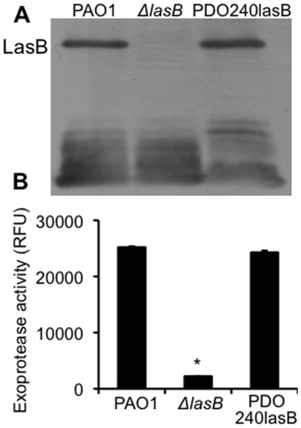
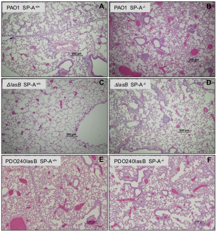
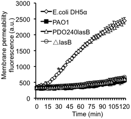
Pseudomonas aeruginosa is an opportunistic pathogen that causes both acute pneumonitis in immunocompromised patients and chronic lung infections in individuals with cystic fibrosis and other bronchiectasis. Over 75% of clinical isolates of P. aeruginosa secrete elastase B (LasB), an elastolytic metalloproteinase that is encoded by the lasB gene. Previously, in vitro studies have demonstrated that LasB degrades a number of components in both the innate and adaptive immune systems. These include surfactant proteins, antibacterial peptides, cytokines, chemokines and immunoglobulins. However, the contribution of LasB to lung infection by P. aeruginosa and to inactivation of pulmonary innate immunity in vivo needs more clarification. In this study, we examined the mechanisms underlying enhanced clearance of the ΔlasB mutant in mouse lungs. The ΔlasB mutant was attenuated in virulence when compared to the wild-type strain PAO1 during lung infection in SP-A+/+ mice. However, the ΔlasB mutant was as virulent as PAO1 in the lungs of SP-A⁻/⁻ mice. Detailed analysis showed that the ΔlasB mutant was more susceptible to SP-A-mediated opsonization but not membrane permeabilization. In vitro and in vivo phagocytosis experiments revealed that SP-A augmented the phagocytosis of ΔlasB mutant bacteria more efficiently than the isogenic wild-type PAO1. The ΔlasB mutant was found to have a severely reduced ability to degrade SP-A, consequently making it unable to evade opsonization by the collectin during phagocytosis. These results suggest that P. aeruginosa LasB protects against SP-A-mediated opsonization by degrading the collectin.


Conflict of interest statement

Competing Interests: The authors have declared that no competing interests exist.

Figures

Figure 1
Figure 1. The ΔlasB mutant has severely attenuated exoprotease activity.
(A) Western blot analysis of the LasB production in the supernatant of PAO1, ΔlasB mutant and PDO240LasB as detected by anti-LasB antibody. (B) Proteolytic activity of stationary phase culture supernatant collected from PAO1, ΔlasB and PDO240lasB. Experiments were performed independently three times in triplicates. The mean + standard deviation from one representative experiment is shown. *p<0.01 when comparing the exoprotease activity of ΔlasB against PAO1 or PDO240lasB.
Figure 2
Figure 2. The ΔlasB mutant is attenuated for virulence in SP-A+/+ mice.
(A) Respiratory tract infections with wild-type PAO1, ΔlasB mutant or genetically-complemented PDO240lasB bacteria were performed by intranasal inoculation of anesthetized SP-A+/+ or SP-A-/- mice. Mouse lungs were harvested 18 hr after infection for CFU enumeration. Data are the mean CFU ± SE (n = 5 per group). * p<0.05 when comparing lungs of SP-A+/+ mice infected with PAO1 and PDO240lasB versus ΔlasB; ** p<0.05 when compared between SP-A+/+ and SP-A-/- mice infected with PAO1, ΔlasB or PDO240lasB bacteria. (B) Mouse lungs were harvested 36 hr after infection for CFU enumeration. Data are the mean CFU ± SE (n = 5 per group). * p<0.05 when comparing lungs of SP-A+/+ mice infected with PAO1 and PDO240lasB versus ΔlasB; ** p<0.05 when compared between SP-A+/+ and SP-A-/- mice infected with PAO1, ΔlasB or PDO240lasB bacteria. (C) Attenuation of ΔlasB bacteria in mouse lungs was not due to a slower growth rate. Bacterial growth was assessed by absorbance at OD600. The data from one of the three independent experiments are shown.
Figure 3
Figure 3. Histopathology of P. aeruginosa infected lungs.
Representative H&E-stained lung sections from SP-A+/+ and SP-A-/- mice (n = 5) 18-hr post intranasal instillation of PAO1 (A-B), ΔlasB (C-D) and PDO240lasB (E-F) bacteria.
Figure 4
Figure 4. SP-A-degrading ability is reduced inΔ lasB mutant bacteria in vitro.
(A) hSP-A (25 µg) was incubated with 1×10⁸ PAO1, ΔlasB or PDO240LasB bacteria for the indicated time intervals. hSP-A degradation was assessed by western blot analyses using 10 µl of SP-A/bacterial suspension. Image from one of the three independent experiments is shown. (B) Densitometry quantification of hSP-A degradation in A. (C) Production of LasB in the mixture was assessed by western blot analyses using 10 µl of supernatant from the hSP-A/bacterial suspension. Immunoblots were probed with anti-SP-A (A) or anti-LasB (C) antibody, respectively.
Figure 5
Figure 5. Elastase deficient ΔlasB mutant is attenuated in the degradation of SP-A during lung infection.
(A-C) The amounts of intact mSP-A were not visibly changed at 6-hr (A) or 12- hr (B) post-infection. By 18 hr post-infection (C), intact mSP-A was reduced in the BAL fluid from PAO1- or PDO240lasB-infected SP-A+/+ mice (n = 6), suggesting that mSP-A was degraded in mouse lungs. In contrast, more abundant mSP-A was clearly visible in the BAL fluids from ΔlasB (n = 8). C  =  Purified human SP-A. M1 – M8  =  BAL of mice infected with P. aeruginosa. Western blot analyses were performed using a polyclonal antibody against SP-A. (D) Densitometry analysis of mSP-A degradation by PAO1, ΔlasB and PDO240lasB in mouse lungs. The amounts of remaining mSP-A in ΔlasB were set to the value of 100%. *p<0.05 when compared the amount of mSP-A in BAL fluids from lungs infected with PAO1 or PDO240lasB against BAL fluids from ΔlasB-infected mice. (E) Mouse BAL from ΔlasB-infected animals contains intact mSP-A that permeabilizes bacterial membranes. Pooled BAL fluids (from C) (50 µg/ml total proteins) were used for membrane permeabilization assays. hSP-A (50 µg/ml) was used as positive control. BAL fluids from PAO1 and PDO240lasB infected mice failed to permeabilize E. coli membranes. hSP-A and BAL samples from ΔlasB-infected mice were able to permeabilize bacterial membranes of E. coli DH5α at higher levels. Experiments were performed independently three times in triplicates. The mean + standard deviation from one representative experiment is shown. **p<0.05 from 60 min onward when comparing the membrane permeabilization of E. coli by pure SP-A or BAL samples from ΔlasB-infected mice against BAL samples from PAO1 or PDO240lasB-infected mice.
Figure 6
Figure 6. ΔlasB mutant bacteria are resistant to SP-A-mediated membrane permeabilization.
Membrane permeabilization assays were performed with 1×10⁸ of E. coli DH5α or P. aeruginosa exposed to hSP-A (50 µg/ml) for 120 min. Three independent experiments were performed in triplicates. The mean + standard deviation from one representative experiment is shown. The membrane permeabilization activity of hSP-A against PAO1, ΔlasB and PDO240lasB was not statistically different among all three P. aeruginosa strains. *p<0.05 from 35 min onward when comparing the membrane permeabilization of DH5α against PAO1, ΔlasB and PDO240lasB.
Figure 7
Figure 7. The ΔlasB mutant is unable to degrade and impede SP-A-mediated opsonization in vitro.
(A) hSP-A opsonized and increased the phagocytosis of both wild-type PAO1 and ΔlasB bacteria in a concentration dependent manner. ∼1×10⁷ PAO1 or ΔlasB bacteria were treated with PBS alone or with increasing concentrations of hSP-A for 1 hr in the presence of 1×10⁶ cultured RAW 264.7 macrophages. The number of phagocytized bacteria was determined by gentamicin exclusion assay. The fold increase in phagocytosis was calculated based on the number of engulfed bacteria in macrophages treated with hSP-A versus PBS alone. Three independent experiments were performed in triplicates. The mean + standard deviation from one representative experiment is shown. *, **p<0.01 when comparing the internalized PAO1 or ΔlasB mutant pretreated with various concentrations of hSP-A versus PBS alone. (B) The ΔlasB mutant bacteria are more susceptible to hSP-A-mediated opsonization. hSP-A (20 µg/ml) was incubated with 1×10⁷ PAO1 or ΔlasB bacteria for 1, 6, 12, or 18 hr. At indicated time intervals, the bacteria-hSP-A mixture was added to 1×10⁶ cultured RAW 264.7 macrophages, and incubated for another 1 hr. The number of engulfed bacteria was examined as in (A), and normalized against PAO1 or ΔlasB bacteria phagocytized in the absence of hSP-A. Three independent experiments were performed in triplicates. The mean + standard deviation from one representative experiment is shown. *p<0.01 when comparing the number of phagocitized ΔlasB bacteria against internalized PAO1 bacteria.
Figure 8
Figure 8. The ΔlasB mutant bacteria are more susceptible to SP-A-mediated opsonization in vivo.
(A) SP-A+/+ mice were intranasally infected with 1×10⁷ of wild-type P. aeruginosa PAO1 or ΔlasB bacteria. At each time interval, infected mice (n = 5) were lavaged for macrophages and infiltrating leukocytes. Cells were centrifuged, washed and the engulfed bacteria were enumerated by gentamicin exclusion assay. Changes in bacterial phagocytosis were calculated based on the number of intracellular PAO1. The mean + standard deviation is shown. *p<0.01 when comparing the number of internalized ΔlasB bacteria against PAO1 bacteria. (B) Leukocyte profiles in mouse lungs infected with PAO1 or ΔlasB bacteria. Macrophages and neutrophils within BAL fluids were determined using antibody specific against each cell type by using flow cytometry. (C) Profiles of macrophage and neutrophil chemotactic chemokines CCL5 and MCP1 in mouse lungs infected with PAO1, ΔlasB or PDO240lasB bacteria.
Figure 9
Figure 9. The ΔlasB mutant bacteria are more susceptible to SP-A-mediated aggregation in vivo.
(A) In vitro aggregation of GFP-expressing wild-type P. aeruginosa PAO1 or ΔlasB bacteria co-incubated with hSP-A and observed under fluorescent microscopy. (B) In vivo aggregation of GFP-expressing wild-type P. aeruginosa PAO1 or ΔlasB (arrows) bacteria lavaged from mouse lungs 18-hr post-infection (n = 5) observed under FLUOVIEW FV300 confocal microscope.
Figure 10
Figure 10. Lysozyme-degrading ability is reduced inΔlasB mutant bacteria in vitro and in vivo.
(A) Chicken lysozyme (5 µg) was incubated with 1×10⁸ PAO1, ΔlasB or PDO240LasB bacteria for the indicated time intervals. Lysozyme degradation was assessed by western blot analyses using 10 µl of lysozyme/bacterial suspension. (B) Elastase deficient ΔlasB mutant is attenuated in the degradation of mouse lysozyme during lung infection. BAL fluids from Fig. 5C were used for western blot analyses. (C) Densitometry quantification of chicken lysozyme degradation in A. (D) Densitometry analysis of lysozyme degradation by PAO1, ΔlasB and PDO240lasB in mouse lungs 18 hr post-infection. The amounts of remaining mouse lysozyme in ΔlasB were set to the value of 100%. *p<0.05 when compared with the amount of lysozyme in BAL fluids from lungs infected with PAO1 or PDO240lasB against BAL fluids from ΔlasB-infected mice.

References

    1. Clements JA. Surface tension of lung extracts. Proc Soc Exp Biol Med. 1957;95:170–172. - PubMed
    1. Crouch E, Wright JR. Surfactant proteins A and D and pulmonary host defense. Annu Rev Physiol. 2001;63:521–554. - PubMed
    1. Hawgood S, Shiffer K. Structures and properties of the surfactant-associated proteins. Annu Rev Physiol. 1991;53:375–394. - PubMed
    1. Holmskov U, Thiel S, Jensenius JC. Collectins and ficolins: humoral lectins of the innate immune defense. Annu Rev Immunol. 2003;21:547–578. - PubMed
    1. Pattle RE. Properties, function and origin of the lining layer. Nature. 1955;175:1125–1126. - PubMed

Publication types

MeSH terms

Substances