Review
. 2010 Nov;2(11):2519-83.
doi: 10.3390/toxins2112519. Epub 2010 Oct 28.

Toxin-based therapeutic approaches

Affiliations
Review

Toxin-based therapeutic approaches

Assaf Shapira et al. Toxins (Basel). 2010 Nov.

Abstract

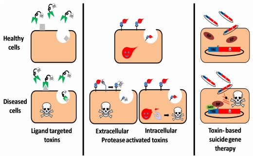
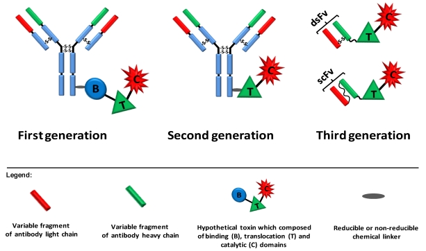
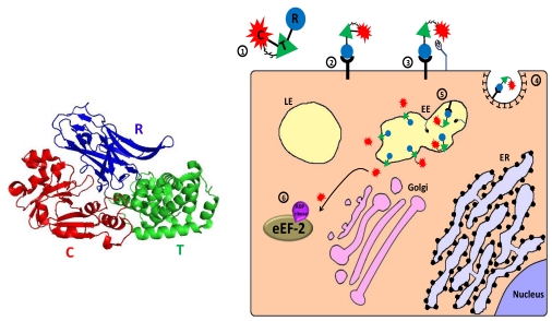
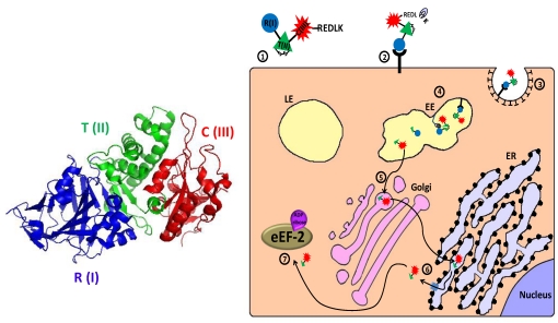
Protein toxins confer a defense against predation/grazing or a superior pathogenic competence upon the producing organism. Such toxins have been perfected through evolution in poisonous animals/plants and pathogenic bacteria. Over the past five decades, a lot of effort has been invested in studying their mechanism of action, the way they contribute to pathogenicity and in the development of antidotes that neutralize their action. In parallel, many research groups turned to explore the pharmaceutical potential of such toxins when they are used to efficiently impair essential cellular processes and/or damage the integrity of their target cells. The following review summarizes major advances in the field of toxin based therapeutics and offers a comprehensive description of the mode of action of each applied toxin.

Keywords: anthrax; diphtheria toxin; immunotoxins; pseudomonas exotoxin A; ricin; suicide gene; targeting; toxins.


Figures

Figure 1
Figure 1
Three targeting strategies in toxin based therapy. Ligand targeted toxins: a ligand (antibody, antibody derivative, cytokine, etc.), which specifically binds to a disease related cell-surface antigen/receptor is linked to a toxic moiety, preferentially as a replacement to the natural cell binding domain of that toxin. Upon administration to patients, the construct selectively binds, is internalized and intoxicates diseased cells, sparing healthy cells that do not display the target on their surface. Protease activated toxins: the toxin is engineered to be cleaved and activated by a disease-related intracellular or extracellular protease. Toxin cleavage may enhance cell-binding and/or translocation, stabilization or catalytic activity of the toxic moiety specifically in protease expressing cells, leading to their eradication. Toxin based suicide gene therapy: a DNA construct, encoding for a toxic polypeptide whose expression is regulated by a specific transcription regulation element (TRE), is delivered to a heterogeneous cell population. However, intoxication occurs only in diseased cells that express an active disease-associated transcription factor (DATF) that specifically binds to the TRE and activates the transcription machinery (RP: RNA polymerase).
Figure 2
Figure 2
Three generations of immunotoxins. First generation immunotoxins were prepared by chemically conjugating antibodies/ligands to intact toxin units or to toxins with attenuated cell binding capability. Reducible or non-reducible chemical bonds/linkers were used for that purpose; the first was generally applied when the conjugation site was positioned on part of the toxin that translocates to the cytosol. In second generation immunotoxins, truncated toxins that lack a cell binding domain were chemically conjugated to a targeting moiety. In third generation immunotoxins, mostly produced in the bacterium Escherichia coli, the cell binding domain of the toxin is genetically replaced with a ligand or with the Fv portion of an antibody in which the light and heavy chain variable fragments are either genetically linked (scFv) or held together by a disulfide bond (dsFv).
Figure 3
Figure 3
Main entry route and mechanism of action of diphtheria toxin. 1. The toxin is secreted as one polypeptide which is composed of three functional domains: the N terminal catalytic domain ((C), also referred to as DTA/DT-A), the translocation domain (T) and the receptor binding domain (R) (see 3D structure (PDB Entry: 1f0l). In the left panel, the colors of the subunits correspond to those in the scheme). In addition, a disulfide bond bridges the C and T domains; 2. The toxin binds via its R domain to a cellular receptor (heparin binding epidermal growth factor precursor); 3. Cell-surface furin protease cleaves the polypeptide chain between the C and T domains that remain linked by a disulfide bond; 4. The toxin-receptor complex is internalized into clathrin coated pits; 5. In the lumen of the early endosome (EE), furin protease cleaves toxin molecules that escaped cell-surface cleavage. The T domain undergoes acidic-induced conformational change, inserted into the endosome membrane and forms a channel through which the catalytic domain can translocate into the cytoplasm where reduction of the interdomain bridging disulfide bond occurs; 6. In the cytoplasm, the catalytic domain inactivates eukaryotic translation elongation factor 2 (eEF2) by ADP-ribosylation, which causes translation inhibition and consequently cell death.
Figure 4
Figure 4
Main entry route and mechanism of action of Pseudomonas exotoxin A. 1. The secreted pseudomonas exotoxin A (PE) toxin can be divided into three main structural and functional domains: the N terminal receptor (R) binding domain I, translocation (T) domain II and the catalytic (C) domain III (see 3D structure (PDB Entry: 1ikq). In the left panel, the colors of the subunits correspond to those in the scheme. For the sake of simplicity, translocation domain II was extended to contain subdomain Ib). A single disulfide bond bridges between cysteines 265 and 287 within domain II; 2. Following removal of a C terminal lysine residue by plasma carboxipeptidase, the toxin binds to its cell-surface receptor (CD91, also called α2MR/LRP); 3. The toxin is internalized mainly via clathrin-coated pits; 4. In the early endosome (EE), the toxin undergoes conformational change and is cleaved by the protease furin in a furin sensitive loop, in domain II. The two cleavage products remain linked by the intradomain disulfide bond; 5. Following reduction of the disulfide bond, the enzymatically active C terminal fragment, which is composed of domain III and about two thirds of domain II, is routed to the trans-Golgi network where it binds via its C terminally exposed REDL sequence to KDEL receptor and travels to the endoplasmic reticulum (ER); 6. In the ER, sequences in domain II mediate the retro‑translocation of the polypeptide via the Sec61p translocon into the cytoplasm; 7. The catalytic domain inactivates eukaryotic translation elongation factor 2 (eEF2) by ADP‑ribosylation, which causes translation inhibition and consequently cell death.
Figure 5
Figure 5
Main entry route and mechanism of action of ricin. 1. Ricin toxin is translated as a single glycosylated polypeptide that is composed of a catalytic A domain and a lectin B domain (see 3D structure (PDB Entry: 2aai) in the left panel; the colors of the subunits correspond to those in the scheme). In the producing plant, a small peptide that links the A and B domains is removed, and the A and B chains remain associated via a single disulfide bond; 2. The toxin binds through the lectin B chain to cell-surface galactose or N‑acetylgalactosamine residues on glycoproteins and glycolipids; 3. Cell-surface bound ricin is internalized by clathrin-dependent as well as clathrin-independent endocytosis and reaches the early endosome (EE); 4. The toxin travels backward through the Golgi to the endoplasmic reticulum (ER), where its’ disulfide linked chains are separated; 5. The catalytic A chain (RTA) is retro-translocated via the Sec61p translocon into the cytoplasm; 6. The catalytically active RTA irreversibly damages ribosome by removing a specific adenine from a conserved 28S rRNA loop (“sarcin/ricin loop”–SRL), which causes translation inhibition and consequently cell death.
Figure 6
Figure 6
Cellular trafficking of anthrax toxin. 1. The toxins are secreted as 3 polypeptides: protective antigen (PA; 83 kDa), lethal factor (LF; 90 kDa) and edema factor (EF; 89 kDa) (see 3D structure (PDB Entry: PA- 1acc; LF-1j7n; EF- 1y0v). In the left panel, the colors of the different proteins correspond to those in the scheme; 2. PA binds to cellular receptor (ATR/TEM8; CMG2); 3. Cell-surface furin protease cleaves PA into an N terminal PA63 (63 kDa) and C terminal PA20 (20 kDa) fragments; 4. Receptor bound PA63 self associates into a homoheptamer (“prepore”) that can bind up to 3 molecules of LF and/or EF; 5. The complex internalized via clathrin-dependent receptor mediated endocytosis; 6. In the early endosomes (EE), the complex is sorted to the vesicular region and preferentially incorporated into intraluminal vesicles. The acidic environment of the endosome induces a conformational change in the prepore that turns into a channel/pore and functions in the translocation of LF and/or EF to the lumen of intraluminal vesicles or to the cytoplasm; 7. Following transportation to late endosome (LE), back fusion of intraluminal vesicles with the limiting membrane delivers the “trapped” toxic factors to the cytoplasm; 8. In the cytoplasm, LF functions as a zinc metalloproteinase that cleaves the N termini of MKK/MEK proteins, blocking their signaling activity. EF acts as a Ca2+/calmodulin activated adenylate cyclase that dramatically elevates cytoplasmic cAMP level and consequently disrupts normal cellular activities.

References

    1. Kostrzewa R., Segura-Aguilar J. Botulinum neurotoxin: Evolution from poison, to research tool—onto medicinal therapeutic and future pharmaceutical panacea. Neurotox. Res. 2007;12:275–290. - PubMed
    1. Mahajan S.T., Brubaker L. Botulinum toxin: From life-threatening disease to novel medical therapy. Am. J. Obstet. Gynecol. 2007;196:7–15. - PubMed
    1. Erbguth F.J. From poison to remedy: The chequered history of botulinum toxin. J. Neur. Transm. 2008;115:559–565. - PubMed
    1. Truong D.D., Stenner A., Reichel G. Current Clinical Applications of Botulinum Toxin. Curr. Pharm. Design. 2009;15:3671–3680. - PubMed
    1. Winau F., Westphal O., Winau R. Paul Ehrlich—In search of the magic bullet. Microb. Infect. 2004;6:786–789. - PubMed