. 2010 Dec;2(12):2824-36.
doi: 10.3390/toxins2122824. Epub 2010 Dec 14.

Gangliosides block Aggregatibacter Actinomycetemcomitans leukotoxin (LtxA)-mediated hemolysis

Affiliations

Gangliosides block Aggregatibacter Actinomycetemcomitans leukotoxin (LtxA)-mediated hemolysis

Michael S Forman et al. Toxins (Basel). 2010 Dec.

Abstract

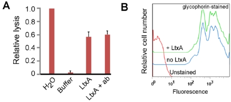
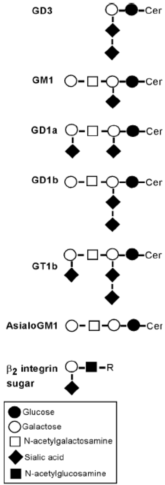
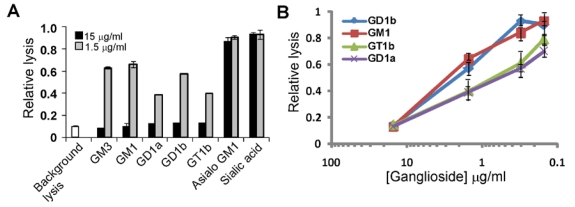
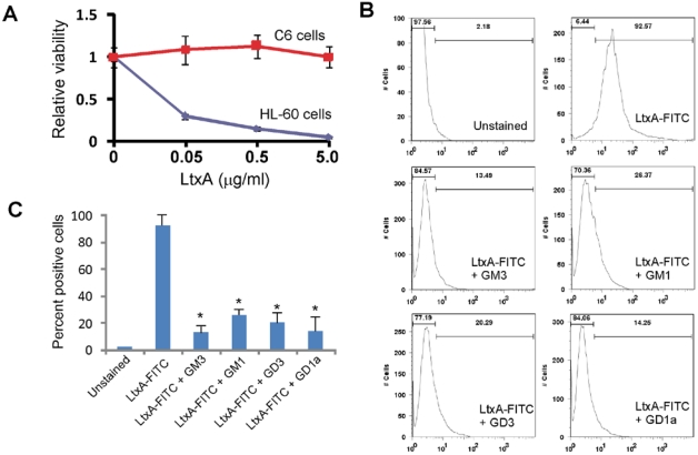
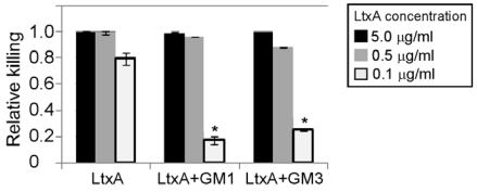
Aggregatibacter actinomycetemcomitans is an oral pathogen and etiologic agent of localized aggressive periodontitis. The bacterium is also a cardiovascular pathogen causing infective endocarditis. A. actinomycetemcomitans produces leukotoxin (LtxA), an important virulence factor that targets white blood cells (WBCs) and plays a role in immune evasion during disease. The functional receptor for LtxA on WBCs is leukocyte function antigen-1 (LFA-1), a β-2 integrin that is modified with N-linked carbohydrates. Interaction between toxin and receptor leads to cell death. We recently discovered that LtxA can also lyse red blood cells (RBCs) and hemolysis may be important for pathogenesis of A. actinomycetemcomitans. In this study, we further investigated how LtxA might recognize and lyse RBCs. We found that, in contrast to a related toxin, E. coli α-hemolysin, LtxA does not recognize glycophorin on RBCs. However, gangliosides were able to completely block LtxA-mediated hemolysis. Furthermore, LtxA did not show a preference for any individual ganglioside. LtxA also bound to ganglioside-rich C6 rat glioma cells, but did not kill them. Interaction between LtxA and C6 cells could be blocked by gangliosides with no apparent specificity. Gangliosides were only partially effective at preventing LtxA-mediated cytotoxicity of WBCs, and the effect was only observed when a high ratio of ganglioside:LtxA was used over a short incubation period. Based on the results presented here, we suggest that because of the similarity between N-linked sugars on LFA-1 and the structures of gangliosides, LtxA may have acquired the ability to lyse RBCs.

Keywords: RTX toxin; endocarditis; erythrocytes; periodontal disease; toxin.


Figures

Figure 1
Figure 1
Effect of glycophorin and LtxA on RBCs. (a) RBCs were treated with water (100% lysis), LtxA buffer (background lysis), LtxA, or first pre-treated with anti-glycophorin antibody (ab) and then LtxA. Lysis was measured by detecting released hemoglobin in the supernatant at 450 nm. The data shown is representative of three independent experiments. (b) Flow cytometry of RBCs stained with anti-glycophorin antibody alone or after pre-treatment with LtxA. The shift in signal to the right represents cells that are stained with anti-glycophorin antibody. The data shown is representative of three independent experiments.
Figure 2
Figure 2
Structures of gangliosides and protein-linked carbohydrates. GD3, GM1, GD1a, GD1b, GT1b, and asialoGM1 are gangliosides. Cer, ceramide; R, complex-type sugar chain as described in [15].
Figure 3
Figure 3
Gangliosides block LtxA-mediated lysis of RBCs. (A) LtxA (7.5 μg/mL) was pre-incubated with sugars (15 μg/mL or 1.5 μg/mL) noted on the x-axis for 20 minutes and then added to RBCs. RBCs were also treated with LtxA buffer to represent background lysis. Experiment was performed in triplicate and standard deviation error bars are shown. GM3, GM1, GD1a, GD1b, and GT1b all caused significant inhibition of LtxA-mediated lysis (P < 0.05). (B) Gangliosides block hemolysis by LtxA in a dose-dependent manner. Results are the average from three independent experiments. LtxA buffer caused relative background lysis of approximately 0.1. All values are relative to LtxA alone (set to 1.0).
Figure 4
Figure 4
Interaction between LtxA-FITC and C6 glioma cells. (A) C6 or HL-60 cells were mixed with varying amounts of LtxA and then incubated for 24 hours. Viability was measured using the CellTiter-Glo viability assay. The experiment was performed in triplicate and standard deviation error bars are shown (P < 0.05). (B) Detection of LtxA-FITC binding to C6 cells using flow cytometry. Cells were stained with LtxA-FITC alone or LtxA-FITC that was pre-incubated (20 minutes) with the ganglioside noted. The left-most bar displays the percent of cells that are negative and the right-most bar represents percent of cells positive for staining with LtxA-FITC. The values on the x-axis represent fluorescence intensity. Results are representative of three experiments. (C) Histogram of the data shown in (B) from three experiments. * represents P < 0.05.
Figure 5
Figure 5
Gangliosides inefficiently block LtxA-mediated toxicity of WBCs. THP-1 cells were treated for three hours with LtxA alone or LtxA pre-incubated with ganglioside GM1 or GM3. The experiment was performed three times and the averages and standard deviations are shown. * represents P < 0.05.

References

    1. Fine D.H., Kaplan J.B., Kachlany S.C., Schreiner H.C. How we got attached to Actinobacillus actinomycetemcomitans: A model for infectious diseases. Periodontol 2000. 2006;42:114–157. doi: 10.1111/j.1600-0757.2006.00189.x. - DOI - PubMed
    1. Slots J., Ting M. Actinobacillus actinomycetemcomitans and Porphyromonas gingivalis in human periodontal disease: Occurrence and treatment. Periodontol 2000. 1999;20:82–121. - PubMed
    1. Zambon J.J. Actinobacillus actinomycetemcomitans in human periodontal disease. J. Clin. Periodontol. 1985;12:1–20. doi: 10.1111/j.1600-051X.1985.tb01348.x. - DOI - PubMed
    1. Lamell C.W., Griffen A.L., McClellan D.L., Leys E.J. Acquisition and colonization stability of Actinobacillus actinomycetemcomitans and Porphyromonas gingivalis in children. J. Clin. Microbiol. 2000;38:1196–1199. - PMC - PubMed
    1. Leys E.J., Griffen A.L., Strong S.J., Fuerst P.A. Detection and strain identification of Actinobacillus actinomycetemcomitans by nested PCR. J. Clin. Microbiol. 1994;32:1288–1294. - PMC - PubMed

Publication types

LinkOut - more resources