

Monitoring radiographic brain tumor progression
- PMID: 22069705
- PMCID: PMC3202817
- DOI: 10.3390/toxins3030191
Monitoring radiographic brain tumor progression
Abstract
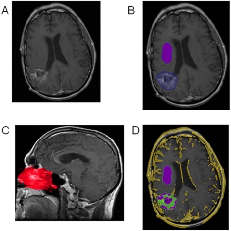
Determining radiographic progression in primary malignant brain tumors has posed a significant challenge to the neuroncology community. Glioblastoma multiforme (GBM, WHO Grade IV) through its inherent heterogeneous enhancement, growth patterns, and irregular nature has been difficult to assess for progression. Our ability to detect tumor progression radiographically remains inadequate. Despite the advanced imaging techniques, detecting tumor progression continues to be a clinical challenge. Here we review the different criteria used to detect tumor progression, and highlight the inherent challenges with detection of progression.
Keywords: MRI; glioblastoma; radiographic progression; tumor progression.
Figures

References
-
- Gururangan S., Krauser J., Watral M.A., Driscoll T., Larrier N., Reardon D.A., Rich J.N., Quinn J.A., Vredenburgh J.J., Desjardins A., et al. Efficacy of high-dose chemotherapy or standard salvage therapy in patients with recurrent medulloblastoma. Neuro. Oncol. 2008;10:745–751. doi: 10.1215/15228517-2008-044. - DOI - PMC - PubMed
-
- Wang S.C., Wikstrom M.G., White D.L., Klaveness J., Holtz E., Rongved P., Moseley M.E., Brasch R.C. Evaluation of Gd-DTPA-labeled dextran as an intravascular MR contrast agent: Imaging characteristics in normal rat tissues. Radiology. 1990;175:483–488. - PubMed
-
- Macdonald D.R., Cascino T.L., Schold S.C., Jr., Cairncross J.G. Response criteria for phase II studies of supratentorial malignant glioma. J. Clin. Oncol. 1990;8:1277–1280. - PubMed
-
- Brandes A.A., Franceschi E., Tosoni A., Blatt V., Pession A., Tallini G., Bertorelle R., Bartolini S., Calbucci F., Andreoli A., et al. MGMT promoter methylation status can predict the incidence and outcome of pseudoprogression after concomitant radiochemotherapy in newly diagnosed glioblastoma patients. J. Clin. Oncol. 2008;26:2192–2197. - PubMed
Publication types
MeSH terms
LinkOut - more resources
Full Text Sources
Other Literature Sources
Medical
