Review
. 2011 Jul;3(7):848-83.
doi: 10.3390/toxins3070848. Epub 2011 Jul 14.

Immunotoxins and anticancer drug conjugate assemblies: the role of the linkage between components

Affiliations
Review

Immunotoxins and anticancer drug conjugate assemblies: the role of the linkage between components

Franco Dosio et al. Toxins (Basel). 2011 Jul.

Abstract

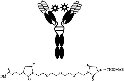
Immunotoxins and antibody-drug conjugates are protein-based drugs combining a target-specific binding domain with a cytotoxic domain. Such compounds are potentially therapeutic against diseases including cancer, and several clinical trials have shown encouraging results. Although the targeted elimination of malignant cells is an elegant concept, there are numerous practical challenges that limit conjugates' therapeutic use, including inefficient cellular uptake, low cytotoxicity, and off-target effects. During the preparation of immunoconjugates by chemical synthesis, the choice of the hinge component joining the two building blocks is of paramount importance: the conjugate must remain stable in vivo but must afford efficient release of the toxic moiety when the target is reached. Vast efforts have been made, and the present article reviews strategies employed in developing immunoconjugates, focusing on the evolution of chemical linkers.

Keywords: antibody drug conjugate; anticancer agents; conjugation process; immunotoxin; linker; toxins.


Figures

Figure 1
Figure 1
Structure of immunotoxins (IT) constructs obtained by chemical (A– intact IgG mAb, B– Fab’ fragment) and genetic engineering (C– Fab fragment, D– Fv fragment) procedures. TX as for toxin or fragment; white rectangles = constant regions, black rectangle = variable region of mAb chains; curvy linkage = peptide bond, –SS– = disulfide bond.
Figure 2
Figure 2
Scheme of reaction for synthesis of ITs. X = reacting group toward amino acid terminus; Y = H or alkyl, aryl group; M and L = leaving groups, stable in buffer but reactive in thiol-disulfide exchange; A and B = chains of RIP II toxins.
Figure 3
Figure 3
Scheme of heterobifunctional linkers used in conjugate preparations MBS, SPDP, SATA, 2-IT (2-iminothiolane) and linkers with improved hindrance around disulfide linkage. SMPT alpha-alkyl derivatives, SulfoNHS-ATMBA (Sulfosuccinimidyl N-[3-(Acetylthio)-3-methylbutyryl-beta-alanine]), and thioimidates AMPT, M-CDPT.
Figure 4
Figure 4
Scheme of preparation of blocked ricin. A triantennary N-linked oligosaccharide, present on glycopeptides from fetuin, is activated with cyanuric chloride (A chain is not shown).
Figure 5
Figure 5
Examples of linker moieties.
Figure 6
Figure 6
Examples of drug-linked conjugates with hydrazo bonds: (A) doxorubicin-mAb conjugate; (B) Vinca alkaloid bridge to a folate targeting moiety.
Figure 7
Figure 7
Some features of structure-activity relationship of Maytansine.
Figure 8
Figure 8
Synthesis of Maytansinoid-mAb conjugates.
Figure 9
Figure 9
Maytansinoid conjugates with improved pharmacokinetics.
Figure 10
Figure 10
Model of metabolism and activation, of maytansine conjugates in a targeted cell. The number is referred to an IC50 value of maytansine derivatives on COLO205 cell line (given as an example).
Figure 11
Figure 11
DM1-mAb conjugate with hydrophilic spacer.
Figure 12
Figure 12
Structure of auristatins (R=CH3) and monomethylauristatins (R=H).
Figure 13
Figure 13
Structure of Auristatin E and MMAE conjugates. The wavy lines indicate the site of hydrolysis (enzymatic or pH-dependent).
Figure 14
Figure 14
ADC composed of a glucuronidase activating linker (1) and the release mechanism.
Figure 15
Figure 15
Structure of Calicheamicin gamma 1.
Figure 16
Figure 16
Mechanism of DNA cleavage by calicheamicin.
Figure 17
Figure 17
Structure of N-acetyl, gamma calicheamicin conjugate: Mylotarg.
Figure 18
Figure 18
Scheme of specific insertion points on thio-trastuzumab and structure of the hydrophilic spacer.

References

    1. Cancer facts and figures. [(accessed on 6 July 2011)]. Available online: http://www.cancer.org/acs/groups/content/@epidemiologysurveilance/docume....
    1. Greish K., Fang J., Inutsuka T., Nagamitsu A., Maeda H. Macromolecular therapeutics: Advantages and prospects with special emphasis on solid tumour targeting. Clin. Pharmacokinet. 2003;42:1089–1105. - PubMed
    1. Maeda H. Tumor-selective delivery of macromolecular drugs via the EPR effect: Background and future prospects. Bioconjugate Chem. 2010;21:797–802. - PubMed
    1. Jain R.K. Transport of molecules across tumor vasculature. Cancer Metastasis Rev. 1987;6:559–593. - PubMed
    1. Roberts W.G., Palade G.E. Neovasculature induced by vascular endothelial growth factor is fenestrated. Cancer Res. 1997;57:765–772. - PubMed

MeSH terms

LinkOut - more resources