. 2012 Mar;12(3):763-71.
doi: 10.1111/j.1600-6143.2011.03846.x. Epub 2011 Nov 9.

B-cell depletion extends the survival of GTKO.hCD46Tg pig heart xenografts in baboons for up to 8 months

Affiliations

B-cell depletion extends the survival of GTKO.hCD46Tg pig heart xenografts in baboons for up to 8 months

M M Mohiuddin et al. Am J Transplant. 2012 Mar.

Abstract

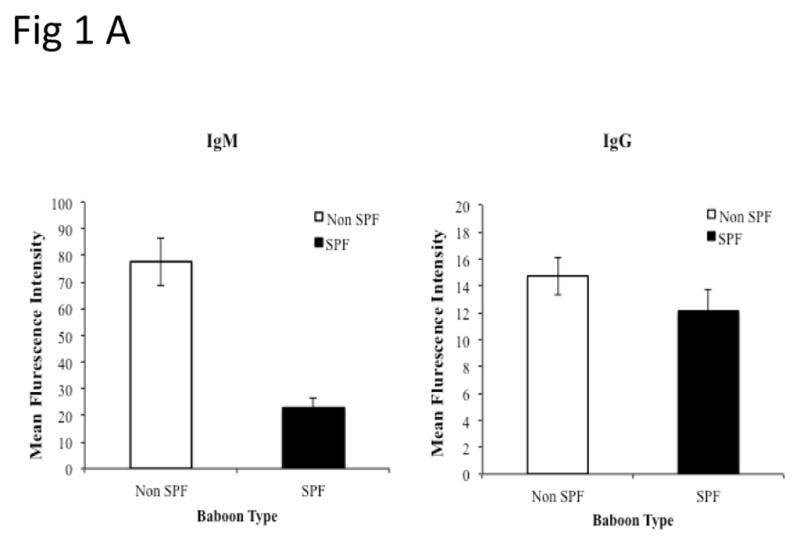
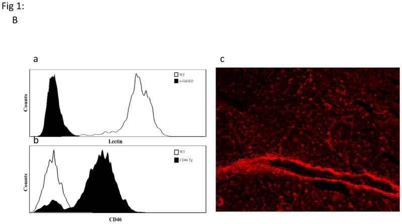
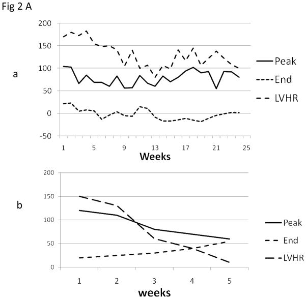
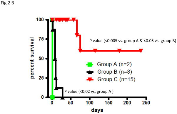
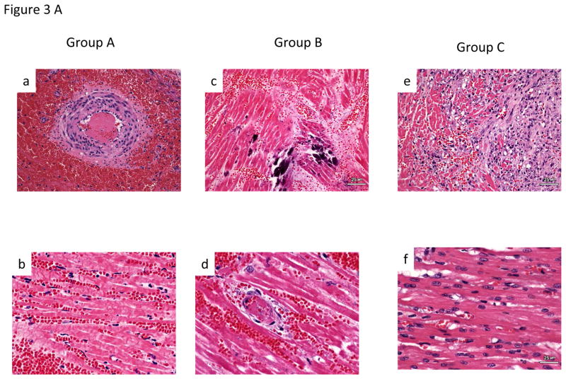
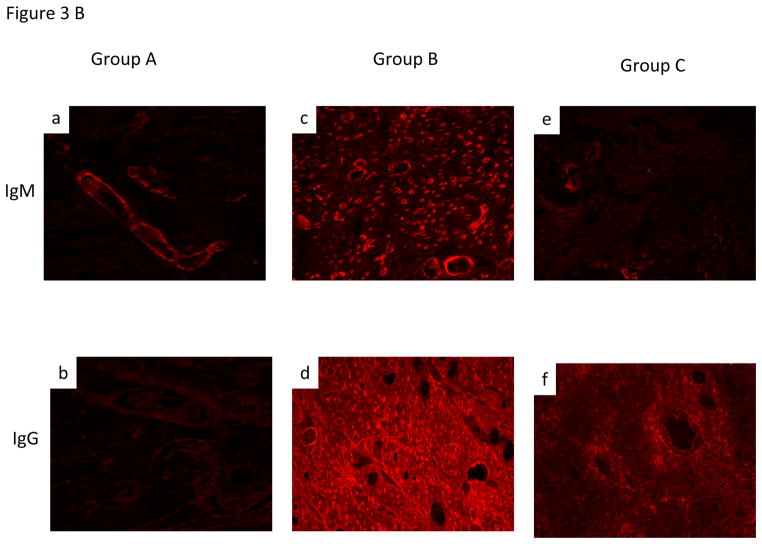
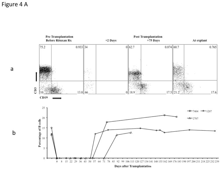
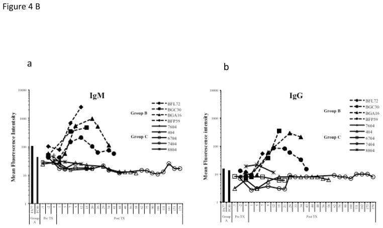
Xenotransplantation of genetically modified pig organs offers great potential to address the shortage of human organs for allotransplantation. Rejection in Gal knockout (GTKO) pigs due to elicited non-Gal antibody response required further genetic modifications of donor pigs and better control of the B-cell response to xenoantigens. We report significant prolongation of heterotopic alpha Galactosyl transferase "knock-out" and human CD46 transgenic (GTKO.hCD46Tg) pig cardiac xenografts survival in specific pathogen free baboons. Peritransplant B-cell depletion using 4 weekly doses of anti-CD20 antibody in the context of an established ATG, anti-CD154 and MMF-based immunosuppressive regimen prolonged GTKO.hCD46Tg graft survival for up to 236 days (n = 9, median survival 71 days and mean survival 94 days). B-cell depletion persisted for over 2 months, and elicited anti-non-Gal antibody production remained suppressed for the duration of graft follow-up. This result identifies a critical role for B cells in the mechanisms of elicited anti-non-Gal antibody and delayed xenograft rejection. Model-related morbidity due to variety of causes was seen in these experiments, suggesting that further therapeutic interventions, including candidate genetic modifications of donor pigs, may be necessary to reduce late morbidity in this model to a clinically manageable level.


Conflict of interest statement

Disclosure:

Two authors of this manuscript have conflicts of interest to disclose as described by the American Journal of Transplantation.

David Ayares is the CEO &president of Revivicor, Inc

Richard Pierson serves on the scientific advisory board of Revivicor, Inc

Figures

Fig 1
Fig 1
Non Gal antibody levels in SPF baboons and verification of pig genetics: PAEC from GTKO pigs were isolated and mixed with baboon sera (1:10 ratio). After incubation, the cells were further stained with PE-goat anti human IgG/IgM or 20 micro L of 1:10 FITC/PE. The mean fluorescence intensity of the cells for each test and control serum was measured for the estimation of non-Gal antibodies. (B) Expression of Gal epitope and hCD46:Peripheral blood lymphocytes of pigs with modified genetics were tested by flow cytometry for the absence of Gal epitope using IB4 lectin (Ba) and transgenic expression of human CD46 (Bb). Expression of hCD46 was confirmed by immunohistochemical staining of heart vessels (Bc).
Fig 1
Fig 1
Non Gal antibody levels in SPF baboons and verification of pig genetics: PAEC from GTKO pigs were isolated and mixed with baboon sera (1:10 ratio). After incubation, the cells were further stained with PE-goat anti human IgG/IgM or 20 micro L of 1:10 FITC/PE. The mean fluorescence intensity of the cells for each test and control serum was measured for the estimation of non-Gal antibodies. (B) Expression of Gal epitope and hCD46:Peripheral blood lymphocytes of pigs with modified genetics were tested by flow cytometry for the absence of Gal epitope using IB4 lectin (Ba) and transgenic expression of human CD46 (Bb). Expression of hCD46 was confirmed by immunohistochemical staining of heart vessels (Bc).
Fig 2
Fig 2. Cardiac xenograft survival and telemetry interpretations
(A): A typical measurement of telemetry readings from one representative baboon is shown in fig 2Aa (Long term survivor) and 2Ab (Rejection within a month) (B): The cardiac xenograft survival in all three groups. In group A and B, grafts were explanted at the time of rejection and data represents the graft survival time while in group C 85% of the recipients died with a functioning xenograft therefore the data points represent the time at which data was censored due to recipient’s death.
Fig 3
Fig 3. Histological and IHC evaluation of the cardiac xenograft
A.Histology: Group A-Section of right ventricle with extensive acute interstitial hemorrhage and arterial thrombus (200X) (A). Section of interventricular septum with diffuse coagulative necrosis and acute interstitial hemorrhage (400X)(B). Group B- (C). Section of left ventricle with focal dystrophic mineralization of myofibers (200X) (C). Section of left ventricular with venular thrombus, coagulative necrosis of adjacent cardiac myocytes and acute interstitial hemorrhage (400X) (D). Group C- Section of left atrium with coagulative necrosis of myocytes, loss of myocytes and replacement of myocytes with moderate interstitial fibrosis with increased numbers of fibroblasts and adjacent collagen (200X)(E). Section of interventricular septum demonstrating an area of relatively normal cardiac myocytes (400X)(F). B. Immunohistochemical staining of graft for non-Gal antibody deposition for both IgM and IgG antibody was consistently observed in all three groups. The stronger intensity of staining in group B likely reflects presence of induced antibodies.
Fig 4
Fig 4. Suppression of B and T cells by the immunosuppression regimen
Analyses of B cell suppression in representative baboons after four doses of anti CD20, at different time points, by flow cytometery is shown (CD19)(4A, a&b) along with suppression of T cells (CD3) by other agents in the immunosuppressive regimen (4Aa). Complete B cell depletion was observed for 60 days with the last dose of anti CD20 given on day 14 post transplantation. Comparison of non Gal (IgM (4Ba) & IgG (4Bb)) antibody mean fluorescent intensity (MFI) of representative baboons rejecting pig hearts; without immunosuppression (Group A)(shown as bars as there was only one time point), immunosuppression without anti CD20 (Group B), and with full immunosuppression including anti CD20 (Group C).

Similar articles

Cited by

References

    1. Mulligan MS, Shearon TH, Weill D, Pagani FD, Moore J, Murray S. Heart and lung transplantation in the United States, 1997–2006. Am J Transplant. 2008 Apr;8(4 Pt 2):977–87. - PubMed
    1. Badiwala MV, Rao V. Left ventricular device as destination therapy: are we there yet? Curr Opin Cardiol. 2009 Mar;24(2):184–9. - PubMed
    1. Cozzi E, Bosio E, Seveso M, Vadori M, Ancona E. Xenotransplantation-current status and future perspectives. Br Med Bull. 2005;75–76:99–114. - PubMed
    1. Mohiuddin MM. Clinical xenotransplantation of organs: why aren’t we there yet? PLoS Med. 2007 Mar 27;4(3):e75. - PMC - PubMed
    1. Schuurman HJ, Pierson RN., III Progress towards clinical xenotransplantation. Front Biosci. 2008;13:204–20. - PubMed

Substances