. 2011 Sep;205(3):265.e1-8.
doi: 10.1016/j.ajog.2011.06.037. Epub 2011 Jun 17.

Molecular phenotype of monocytes at the maternal-fetal interface

Affiliations

Molecular phenotype of monocytes at the maternal-fetal interface

Subhabrata Basu et al. Am J Obstet Gynecol. 2011 Sep.

Abstract

Objective: The purpose of this study was to gain insight into the pathways that are associated with inflammation at the maternal-fetal interface. This study examined the molecular characteristics of monocytes that were derived from the maternal circulation and the placenta of obese women.

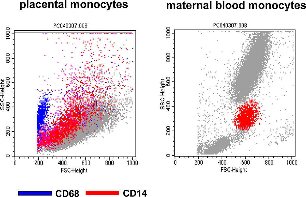
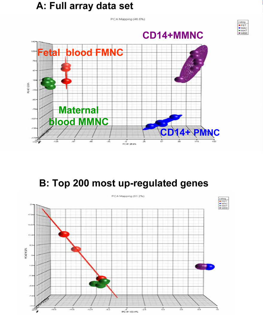
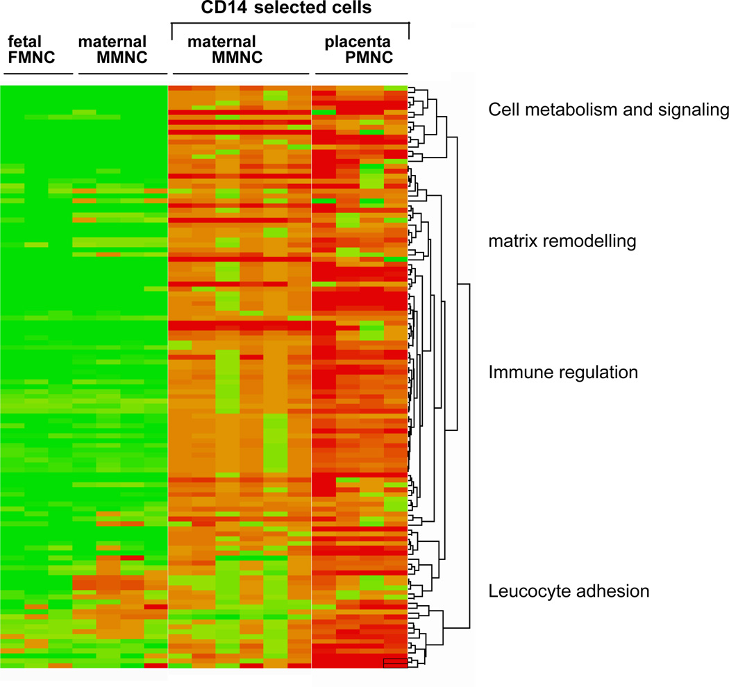
Study design: Mononuclear cells were isolated from placenta, venous maternal, and umbilical cord blood at term delivery; activated monocytes were separated with CD14 immunoselection. The genotype and expression pattern of the monocytes were analyzed by microarray and real-time reverse transcriptase-polymerase chain reaction.

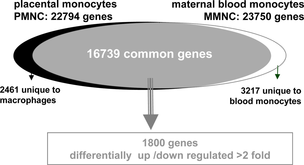
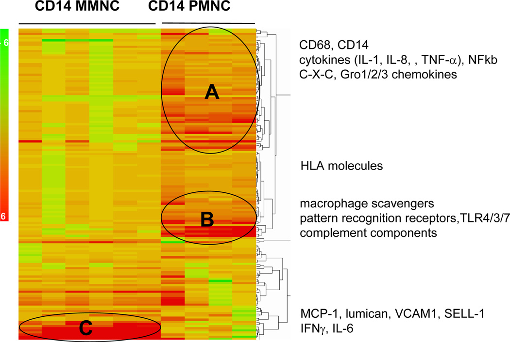
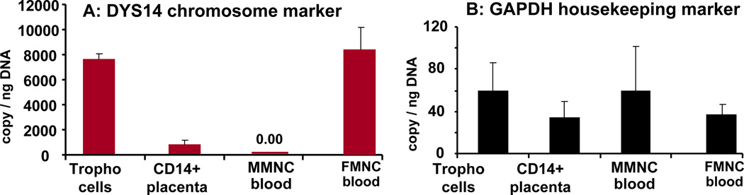
Results: The transcriptome of the maternal blood and placental CD14 monocytes exhibited 73% homology, with 10% (1800 common genes) differentially expressed. Genes for immune sensing and regulation, matrix remodeling, and lipid metabolism were enhanced 2-2006 fold in placenta, compared with maternal monocytes. The CD14 placental monocytes exhibited a maternal genotype (9% DYS14 expression) as opposed to the fetal genotype (90% DYS14 expression) of the trophoblast cells.

Conclusion: CD14 monocytes from the maternal blood and the placenta share strong phenotypic and genotypic similarities with an enhanced inflammatory pattern in the placenta. The functional traits of the CD14 blood and placental monocytes suggest that they both contribute to propagation of inflammation at the maternal-fetal interface.


Conflict of interest statement

DISCLOSURE: The authors report no conflict of interest

Figures

Figure 1
Figure 1. global pattern of gene expression of monocytes from maternal blood and the placenta
The whole transcriptome of CD14 monocytes from placenta (22794 genes) and maternal blood (23750 genes) was obtained by screening oligonucleotide arrays containing a total of 47,000 transcripts. Filtering of raw data identified 16739 genes common to the 2 data sets. 1800 genes having a false discovery rate <0.05 and a fold change >2 were considered differentially expressed between groups.
Figure 2
Figure 2. Flow cytometry analysis of isolated placental and maternal monocytes
Representative double color fluorescent-activated cell sorter analysis using CD14 - PE and CD68-FITC labeled antibodies. Left panel : dot plot side scattered (SSC) of isolated placental double staining with CD14 and CD68 shows wide distribution and size of CD14 positive cells. Right panel: double staining of maternal mononuclear blood cells. Six cell preparations from independent placentas and blood of women with BMI >25 were analyzed.
Figure 3
Figure 3. principal component analysis (PCA) of the systemic mononuclear cells and placental macrophages
Three dimensional scatter plots PCA analysis of the transcriptome of CD14 immnuno-selected monocytes from maternal blood (MMNC) and the placenta (PMNC) and non immuno-selected monocytes from maternal and umbilical blood. A: Analysis of full data set shows a segregation pattern between the CD14 immunoselected MMNC and PMNC cells and the non immunoselected cells (mat-MNC and fet-MNC) closely grouped together. B: Analysis of the 200 top regulated genes showed the expression trends in MMNC and PMNC are superimposable. Each knot represents one microarray with a pool of independent cell samples obtained from 4–6 subjects.
Figure 4
Figure 4. Hierarchical clustering of the top 200 most induced genes in placental macrophages of obese women
Clustering generated heat map identified four primary functional clusters based on biological annotation of the genes up-regulated in CD14 immunoselected compared to unselected monocytes cells from maternal and cord blood.
Figure 5
Figure 5. Differential expression of genes regulating immune response in monocytes of maternal blood and the placenta
GO annotation of immune function and pathways studio significantly up-regulated identified 150 genes based on p values < 10.E−5.
Figure 6
Figure 6. gender characterization of tissue resident macrophages
Expression of DYS14 markers for the Y chromosome was quantitated by RT-PCR. PMNC of male fetuses exhibited a low Y copy numbers (p<0.001) compared to cord blood mononuclear cells. The level of Y expression was undetectable in MMNC of maternal blood. The expression of the house keeping gene glyceraldehyde -3-P(GAPDH) is gender independent i.e. same level in maternal and fetal cells. Results are means ± SEM of 6 independent samples from male fetuses with determinations performed in duplicate. All values were normalized for actin.

References

    1. Renaud SJ, Graham CH. The role of macrophages in utero-placental interactions during normal and pathological pregnancy. Immunol Invest. 2008;37:535–564. - PubMed
    1. Szekeres-Bartho J. Immunological relationship between the mother and the fetus. Int Rev Immunol. 2002;21:471–495. - PubMed
    1. Challis JR, Lockwood CJ, Myatt L, Norman JE, Strauss JF, 3rd, Petraglia F. Inflammation and pregnancy. Reprod Sci. 2009;16:206–215. - PubMed
    1. Kim JS, Romero R, Cushenberry E, Kim YM, Erez O, Nien JK, Yoon BH, Espinoza J, Kim CJ, Dennis G. Distribution of CD14+ and CD68+ macrophages in the placental bed and basal plate of women with preeclampsia and preterm labor. Placenta. 2007;5–6:571–576. - PubMed
    1. Challier JC, Bintein T, Basu S, Hotmire K, Minium J, Catalano PM, et al. Obesity in Pregnancy stimulates macrophage accumulation and inflammation in the placenta. Placenta. 2008;29:274–281. - PMC - PubMed

Publication types

Substances