. 2012 Mar;5(2):241-7.
doi: 10.1242/dmm.008326. Epub 2011 Nov 9.

Haploinsufficiency of the genes encoding the tumor suppressor Pten predisposes zebrafish to hemangiosarcoma

Affiliations

Haploinsufficiency of the genes encoding the tumor suppressor Pten predisposes zebrafish to hemangiosarcoma

Suma Choorapoikayil et al. Dis Model Mech. 2012 Mar.

Abstract

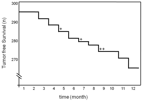
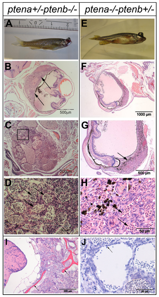
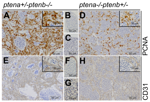
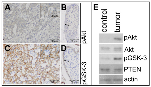
PTEN is an essential tumor suppressor that antagonizes Akt/PKB signaling. The zebrafish genome encodes two Pten genes, ptena and ptenb. Here, we report that zebrafish mutants that retain a single wild-type copy of ptena or ptenb (ptena(+/-)ptenb(-/-) or ptena(-/-)ptenb(+/-)) are viable and fertile. ptena(+/-)ptenb(-/-) fish develop tumors at a relatively high incidence (10.2%) and most tumors developed close to the eye (26/30). Histopathologically, the tumor masses were associated with the retrobulbar vascular network and diagnosed as hemangiosarcomas. A single tumor was identified in 42 ptena(-/-)ptenb(+/-) fish and was also diagnosed as hemangiosarcoma. Immunohistochemistry indicated that the tumor cells in ptena(+/-)ptenb(-/-) and ptena(-/-)ptenb(+/-) fish proliferated rapidly and were of endothelial origin. Akt/PKB signaling was activated in the tumors, whereas Ptena was still detected in tumor tissue from ptena(+/-)ptenb(-/-) zebrafish. We conclude that haploinsufficiency of the genes encoding Pten predisposes to hemangiosarcoma in zebrafish.


Figures

Fig. 1.
Fig. 1.
Ocular tumor incidence in ptena+/−ptenb−/− zebrafish over time. Kaplan-Meier plot of tumor incidence. Ptena+/−ptenb−/− zebrafish (n=294) were monitored over time. Number of tumor-free fish are plotted on the y-axis (n). Most tumors (26/30) developed close to the eye. The tumors in other locations (4/30) are indicated as +.
Fig. 2.
Fig. 2.
Ocular tumor of ptena+/−ptenb−/− and ptena−/−ptenb+/− mutants, diagnosed as hemangiosarcoma. (A–D) 3-month-old ptena+/−ptenb−/− and (E-H) 9-month-old ptena−/−ptenb+/− mutant with ocular tumor. The entire intact fish was fixed and embedded in paraffin. (B–D) Transversal sections and (F–H) sagittal sections were stained with H&E. Arrows indicate tumor mass, which is associated with the eye bulbs. (B,G) Higher-power magnifications of the tumor mass; (D) magnification of the boxed area in C. The tumor consists of cells that form different sizes of blood-filled spaces (arrows in D,H).(I,J) H&E staining of sections from two individuals revealed hemangiosarcoma formation. (I) The tumor was invasive and penetrated into the brain region with enclosing scull elements (arrows). (J) Cells with plump morphology (arrow) are detaching from surrounding tissue and protrude into the vessel lumen. Sections of representative tumors are depicted here.
Fig. 3.
Fig. 3.
Low-dose Pten tumors display elevated cell proliferation and have endothelial cell origin. ptena+/−ptenb−/− (n=6) (A–C,E–G) and ptena−/−ptenb+/− (n=1) (D,H) mutants with tumors were fixed, paraffin embedded and sectioned transversally or sagitally. Immunohistochemistry using PCNA, a cell proliferation marker, showed clearly enhanced nuclear PCNA staining in tumor cells (A,D) compared with control tissue in the same sections (B,C). CD31, an endothelial cell marker, was expressed in the tumor tissue (E,H) in a similar manner as in control blood vessels in the same sections (F,G). Representative sections are depicted here.
Fig. 4.
Fig. 4.
Elevated Akt/PKB signaling in Pten mutant tumors. Immunohistochemistry on transversal sections from a ptena+/−ptenb−/− mutant fish, using pAkt- and pGSK-3β-specific antibodies. (A) Tumor area stains weakly positive for pAkt, whereas (B) control vessel (arrow) from the same section is not stained (n=10). (C) Tumor area is highly positive for pGSK-3β, whereas (D) cells from a control vessel (arrow) from the same section only stain mildly positive (n=6). (E) The cranial part of a ptena+/−ptenb−/− mutant that developed a tumor was dissected into two fragments, one harboring tumor tissue (tumor) and the other representing control tissue (control). The samples were lysed and the lysates were run on a denaturing SDS-polyacrylamide gel. The proteins were transferred to a PVDF membrane and after blocking the blot was probed with anti-pAkt antibody, stripped and sequentially probed with anti-Akt, anti-pGSK-3, anti-PTEN and, as a loading control, anti-actin.

References

    1. Ali I. U., Schriml L. M., Dean M. (1999). Mutational spectra of PTEN/MMAC1 gene: a tumor suppressor with lipid phosphatase activity. J. Natl. Cancer Inst. 91, 1922–1932 - PubMed
    1. Alimonti A., Carracedo A., Clohessy J. G., Trotman L. C., Nardella C., Egia A., Salmena L., Sampieri K., Haveman W. J., Brogi E., et al. (2010). Subtle variations in Pten dose determine cancer susceptibility. Nat. Genet. 42, 454–458 - PMC - PubMed
    1. Barnett C. H. (1951). The structure and function of the choroidal gland of teleostean fish. J. Anat. 85, 113–119 - PMC - PubMed
    1. Berghmans S., Murphey R. D., Wienholds E., Neuberg D., Kutok J. L., Fletcher C. D., Morris J. P., Liu T. X., Schulte-Merker S., Kanki J. P., et al. (2005). tp53 mutant zebrafish develop malignant peripheral nerve sheath tumors. Proc. Natl. Acad. Sci. USA 102, 407–412 - PMC - PubMed
    1. Biggs W. H., 3rd, Meisenhelder J., Hunter T., Cavenee W. K., Arden K. C. (1999). Protein kinase B/Akt-mediated phosphorylation promotes nuclear exclusion of the winged helix transcription factor FKHR1. Proc. Natl. Acad. Sci. USA 96, 7421–7426 - PMC - PubMed

Publication types

MeSH terms

Substances

LinkOut - more resources