

High tibial osteotomy in medial compartment osteoarthritis and varus deformity using the Taylor spatial frame: early results
- PMID: 22072322
- PMCID: PMC3225572
- DOI: 10.1007/s11751-011-0123-2
High tibial osteotomy in medial compartment osteoarthritis and varus deformity using the Taylor spatial frame: early results
Abstract
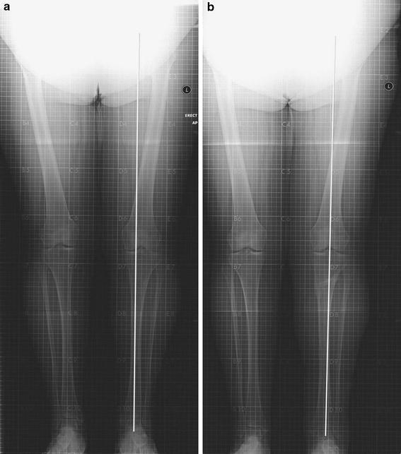
We report the early results of high tibial osteotomy (HTO) in medial compartment osteoarthritis (OA) and varus deformity using the Taylor spatial frame (TSF). Between October 2005 and April 2007, 9 patients with medial compartment OA and varus deformity underwent TSF application and medial opening wedge HTO. Pre- and post-operative Oxford knee scores, SF-12 and visual analogue pain scores were recorded along with radiographic outcomes. Median follow-up was 19 months (range 15-35). Mean age at operation was 49 years (range 37-59). The median time spent in the frame was 18 weeks (range 12-37). The mean preoperative Oxford knee score was 28.7. This improved to a mean of 35.4 post-operatively (P = 0.0142). 6 (67%) patients had a documented pin-site infection. With TKR as an end point, the survival rate of HTOs was 88.9% at a median of 19 months follow-up. This study demonstrates that in selected patients the TSF provides a viable treatment option for performing HTO in medial compartment OA with varus deformity.
Figures

Similar articles
-
High tibial osteotomy with use of the Taylor Spatial Frame external fixator for osteoarthritis of the knee.Can J Surg. 2006 Aug;49(4):245-50. Can J Surg. 2006. PMID: 16948882 Free PMC article.
-
Gait analysis following medial opening-wedge high tibial osteotomy.Knee Surg Sports Traumatol Arthrosc. 2018 Jun;26(6):1838-1844. doi: 10.1007/s00167-017-4421-1. Epub 2017 Mar 1. Knee Surg Sports Traumatol Arthrosc. 2018. PMID: 28251263
-
Predictive factors for satisfaction after contemporary unicompartmental knee arthroplasty and high tibial osteotomy in isolated medial femorotibial osteoarthritis.Orthop Traumatol Surg Res. 2019 Feb;105(1):77-83. doi: 10.1016/j.otsr.2018.11.001. Epub 2018 Dec 1. Orthop Traumatol Surg Res. 2019. PMID: 30509622
-
High tibial osteotomy in the ACL-deficient knee with medial compartment osteoarthritis.J Orthop Traumatol. 2016 Sep;17(3):277-85. doi: 10.1007/s10195-016-0413-z. Epub 2016 Jun 29. J Orthop Traumatol. 2016. PMID: 27358200 Free PMC article. Review.
-
A critical appraisal of medial open wedge high tibial osteotomy for knee osteoarthritis.J Clin Orthop Trauma. 2018 Oct-Dec;9(4):300-306. doi: 10.1016/j.jcot.2018.02.004. Epub 2018 Feb 10. J Clin Orthop Trauma. 2018. PMID: 30449975 Free PMC article. Review.
Cited by
-
A Systematic Review of Patient-reported Outcome Measures Used in Circular Frame Fixation.Strategies Trauma Limb Reconstr. 2019 Jan-Apr;14(1):34-44. doi: 10.5005/jp-journals-10080-1413. Strategies Trauma Limb Reconstr. 2019. PMID: 32559266 Free PMC article. Review.
-
Gradual correction of idiopathic genu varum deformity using the Ilizarov technique.Knee Surg Sports Traumatol Arthrosc. 2013 Jul;21(7):1523-9. doi: 10.1007/s00167-012-2074-7. Epub 2012 Jun 4. Knee Surg Sports Traumatol Arthrosc. 2013. PMID: 22660974
-
Management of Osteoarthritis Knee by Graduated Open Wedge High Tibial Osteotomy in 40-60 Years Age Group Using Limb Reconstruction System: A Clinical Study.J Clin Diagn Res. 2015 Oct;9(10):RC09-11. doi: 10.7860/JCDR/2015/14469.6644. Epub 2015 Oct 1. J Clin Diagn Res. 2015. PMID: 26557580 Free PMC article.
-
Amputation vs Reconstruction in Type IV Tibial Hemimelia: Functional Outcomes and Description of a Novel Surgical Technique.Strategies Trauma Limb Reconstr. 2023 Jan-Apr;18(1):32-36. doi: 10.5005/jp-journals-10080-1576. Strategies Trauma Limb Reconstr. 2023. PMID: 38033924 Free PMC article.
-
Opening Wedge High Tibial Osteotomy with Combined Use of Patient-Specific 3D-Printed Plates and Taylor Spatial Frame for the Treatment of Knee Osteoarthritis.Pain Res Manag. 2021 Dec 1;2021:8609921. doi: 10.1155/2021/8609921. eCollection 2021. Pain Res Manag. 2021. PMID: 34900072 Free PMC article.
References
-
- Jackson JP. Osteotomy for osteoarthritis of the knee. In proceedings of the Sheffield regional orthopaedic club. J Bone Joint Surg Br. 1958;40-B:826.
-
- Coventry MB. Osteotomy of the upper portion of the tibia for degenerative arthritis of the knee. A preliminary report. J Bone Joint Surg Am. 1965;47-A:984. - PubMed
-
- Bauer GCH, Insall JN, Koshino T. Tibial osteotomy in gonarthrosis (osteoarthritis of the knee) J Bone Joint Surg Am. 1969;51-A:1545–1563. - PubMed
-
- Maquet P, de Marchin P, Simonet J. Biom canique du genou et gonarthrose. Rhumatologie. 1967;19:51–70. - PubMed
-
- Coventry MB, Ilstrup DM, Wallrichs SL. Proximal tibial osteotomy. A critical long-term study of eighty-seven cases. J Bone Joint Surg Am. 1993;75-A:196–201. - PubMed
LinkOut - more resources
Full Text Sources
Miscellaneous
