

Quantifying vaginal tissue elasticity under normal and prolapse conditions by tactile imaging
- PMID: 22072417
- PMCID: PMC3306492
- DOI: 10.1007/s00192-011-1592-z
Quantifying vaginal tissue elasticity under normal and prolapse conditions by tactile imaging
Abstract
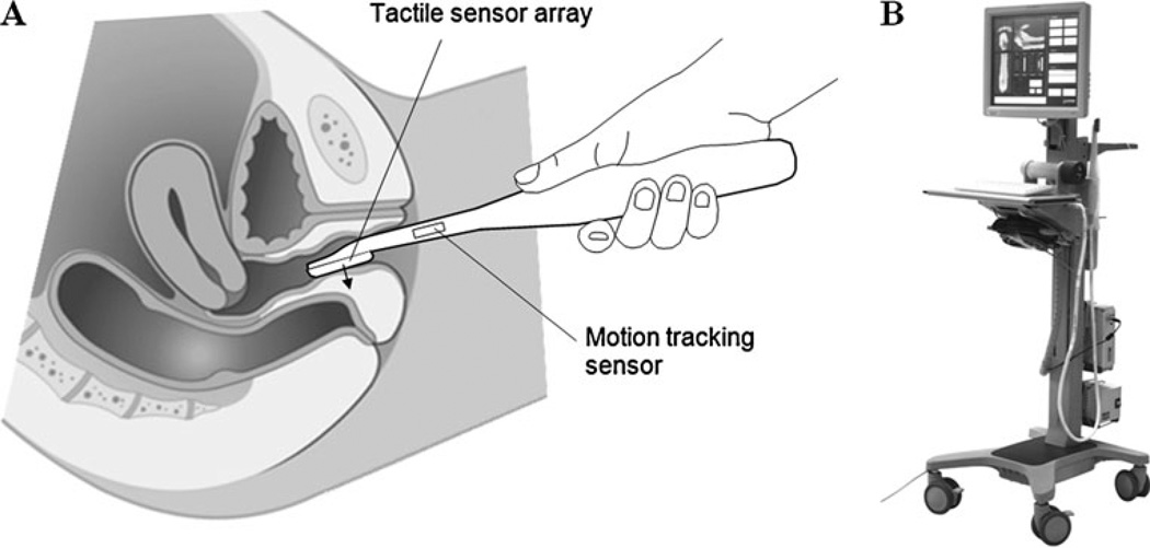
Introduction and hypothesis: Vaginal tactile imaging (VTI) is based on principles similar to those of manual palpation. The objective of this study is to assess the clinical suitability of new approach for imaging and tissue elasticity quantification under normal and prolapse conditions.
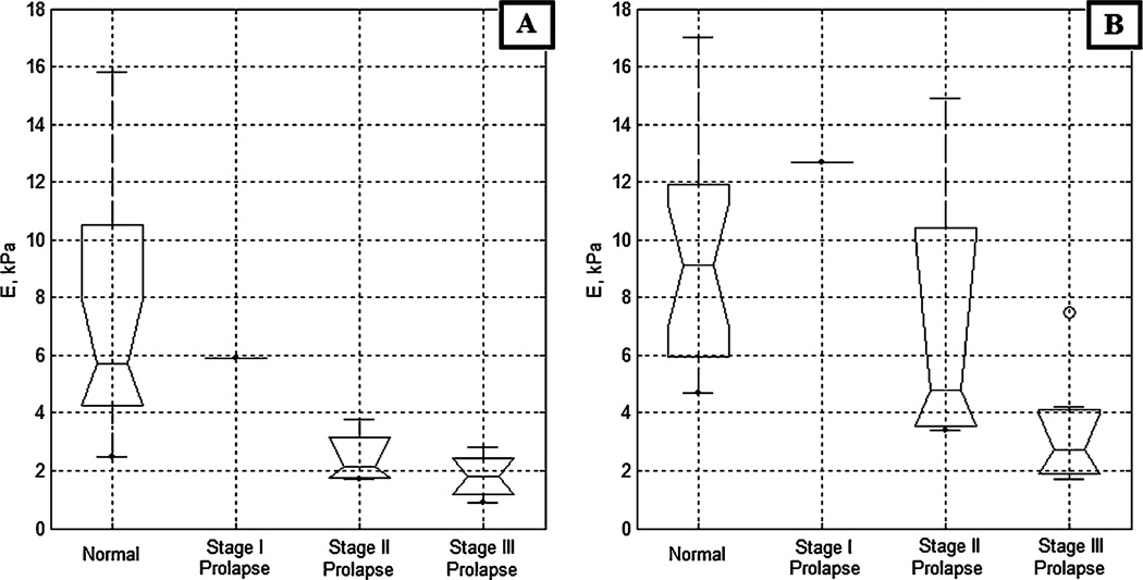
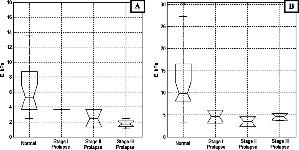
Methods: The study subjects included 31 women with normal and prolapse conditions. The tissue elasticity (Young's modulus) was calculated from spatial gradients in the resulting 3-D tactile images.
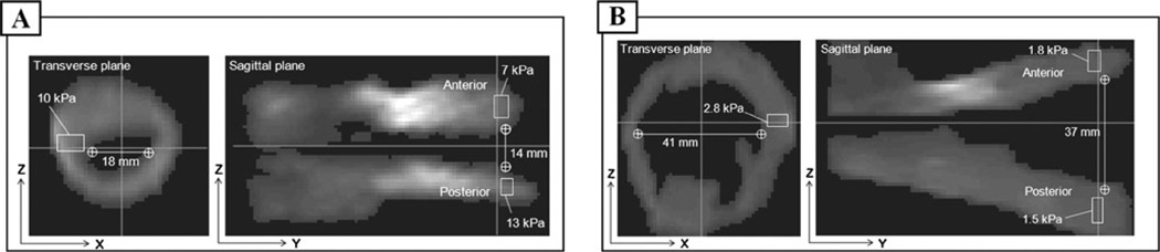
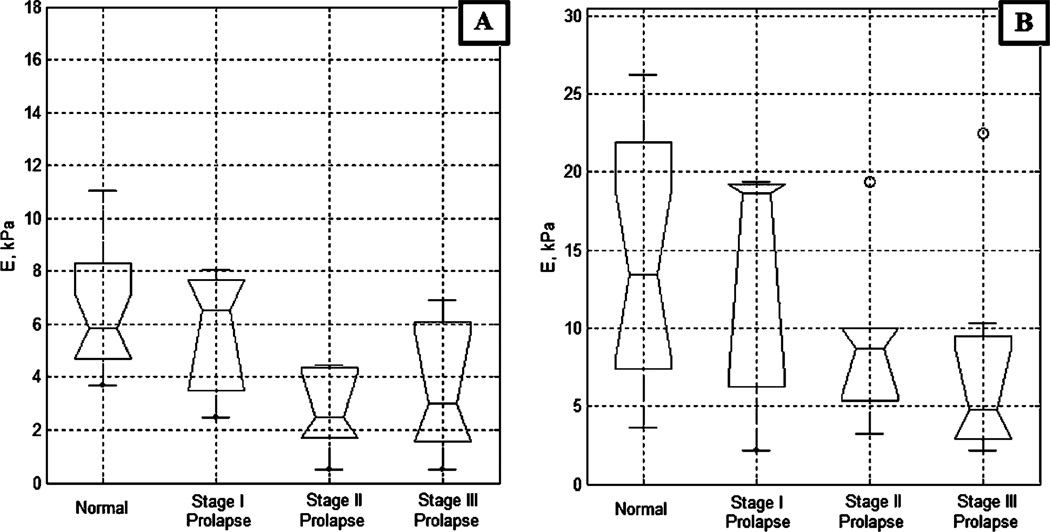
Results: Average values for tissue elasticity for the anterior and posterior compartments for normal conditions were 7.4 ± 4.3 kPa and 6.2 ± 3.1 kPa respectively. For Stage III prolapse the average values for tissue elasticity for anterior and posterior compartments were 1.8 ± 0.7 kPa and 1.8 ± 0.5 kPa respectively.
Conclusions: VTI may serve as a means for 3-D imaging of the vagina and a quantitative assessment of vaginal tissue elasticity, providing important information for furthering our understanding of pelvic organ prolapse and surgical treatment.
Conflict of interest statement
Figures
References
-
- Swift SE. The distribution of pelvic organ support in a population of female subjects seen for routine gynecologic health care. Am J Obstet Gynecol. 2000;183:277–285. - PubMed
-
- Jelovsek JE, Maher C, Barber MD. Pelvic organ prolapse. Lancet. 2007;369:1027–1038. - PubMed
-
- Abramowitch SD, Feola A, Jallah Z, Moalli PA. Tissue mechanics, animal models, and pelvic organ prolapse: a review. Eur J Obstet Gynecol Reprod Biol. 2009;144:S146–S158. - PubMed
-
- Jean-Charles C, Rubod C, Brieu M, Boukerrou M, Fasel J, Cosson M. Biomechanical properties of prolapsed or non-prolapsed vaginal tissue: impact on genital prolapse surgery. Int Urogynecol J. 2010;21:1535–1538. - PubMed
-
- Ophir J, Cespedes I, Ponnekanti H, Yazdi Y, Li X. Elastography: a quantitative method for imaging the elasticity of biological tissues. Ultrason Imaging. 1991;13:111–134. - PubMed
Publication types
MeSH terms
Grants and funding
LinkOut - more resources
Full Text Sources
Medical
