Review
. 2011 Nov 9;31(45):16076-85.
doi: 10.1523/JNEUROSCI.3454-11.2011.

The neurophysiology and pathology of brain zinc

Affiliations
Review

The neurophysiology and pathology of brain zinc

Stefano L Sensi et al. J Neurosci. .

Abstract

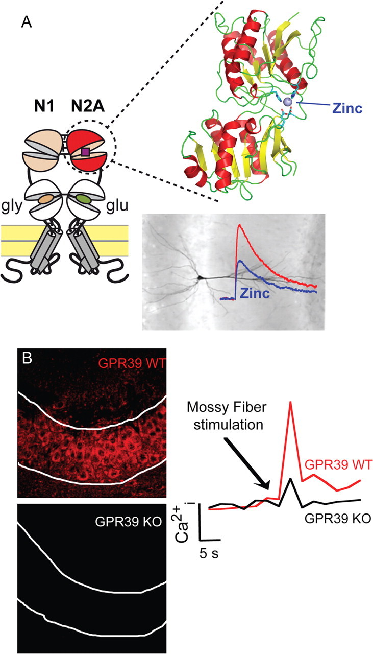
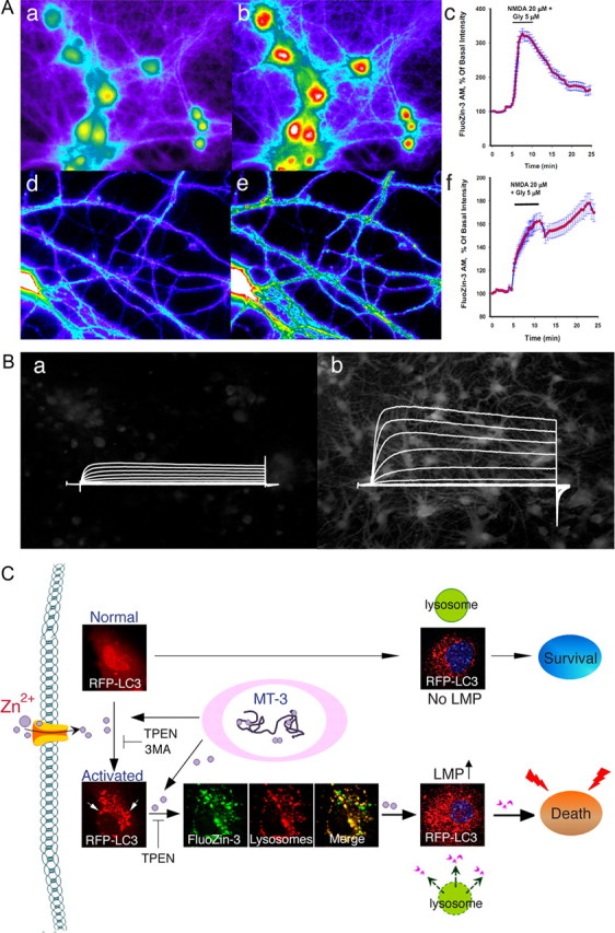
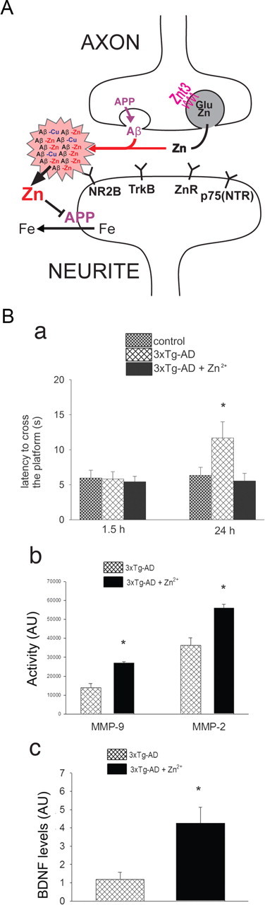
Our understanding of the roles played by zinc in the physiological and pathological functioning of the brain is rapidly expanding. The increased availability of genetically modified animal models, selective zinc-sensitive fluorescent probes, and novel chelators is producing a remarkable body of exciting new data that clearly establishes this metal ion as a key modulator of intracellular and intercellular neuronal signaling. In this Mini-Symposium, we will review and discuss the most recent findings that link zinc to synaptic function as well as the injurious effects of zinc dyshomeostasis within the context of neuronal death associated with major human neurological disorders, including stroke, epilepsy, and Alzheimer's disease.


Figures

Figure 1.
Figure 1.
Zn2+ and synaptic function. A, A high-affinity “Zn2+ sensor” in NMDA receptors. NMDAR subunits contain in their extracellular region a tandem of clamshell-like domains, the N-terminal domain (NTD) and the agonist-binding domain, which is directly connected to the transmembrane pore region. In the GluN2A subunit, the NTD forms a discrete high-affinity Zn2+ binding site that underlies allosteric inhibition of NMDAR-mediated synaptic currents by nanomolar Zn2+ concentrations. B, Synaptic Zn2+ activates a specific mZnR. Synaptic Zn2+ released from the mossy fibers activates metabotropic Ca2+ release via the ZnR. The expression of GPR39 (left panel) and synaptic Zn2+-dependent Ca2+ release (right panel) are eliminated in the CA3 pyramidal cell layer in GPR39 KO mice. The activity of the mZnR triggers phosphorylation of ERK1/2 and regulation of Cl transport, which lead to increased inhibitory drive.
Figure 2.
Figure 2.
Zn2+-mediated neuronal death and dysfunction. A, NMDAR activation induces [Zn2+]i mobilization in neuronal soma and dendrites. Neurons loaded with the Zn2+-selective probe FluoZin3-AM (a, d) were exposed to NMDA (20 μm) for 5 min. NMDAR activation induces [Zn2+]i rises (b, e) that show a different profile when analyzing changes in neuronal somata (c) compared to dendrites (f). Time course graphs show changes in fluorescence levels in neuronal somata and dendrites before and after the NMDA challenge, respectively. Note how the dendritic [Zn2+]i rises are long lasting, while in the soma, [Zn2+]i levels rapidly recover to baseline. B, A Zn2+–K+ continuum in neuronal apoptosis. Oxidant exposure in neurons (a) results in the liberation of Zn2+ from intracellular metal binding proteins (b). This Zn2+ activates a signaling cascade that ultimately produces a robust enhancement of voltage-activated delayed rectifier K+ currents (b). This, in turn, leads to the loss of intracellular K+, creating a permissive environment for the completion of apoptotic programs. C, Zn2+ influx or intracellular Zn2+ release from metallothionein 3 (MT-3) activates autophagy and causes accumulation of Zn2+ in autophagosomes and autolysosomes. Under physiological conditions, activated autophagy serves beneficial functions by removing abnormal proteins and organelles. However, when in excess, it leads to LMP and neuronal death.
Figure 3.
Figure 3.
Zn2+ in Alzheimer's disease. A, Zn2+ released during neurotransmission is trapped by amyloid, depriving targets essential for LTP. In addition, the Zn2+ transfers inappropriately to APP and inhibits its ferroxidase activity and ability to facilitate iron release from neurons, leading to pro-oxidant intraneuronal iron accumulation as a downstream consequence of extracellular Zn2+ accumulation. B, Zn2+ supplementation is beneficial in an animal model of AD. 3xTg-AD mice chronically fed (11–13 months) with water containing 30 ppm of ZnSO4 are protected from the appearance at 12–14 months of age of hippocampus-dependent memory deficits (as assessed with the Morris water maze test). Mice were tested when the platform was removed 1.5 h (a, left panel; to investigate short-term memory) and 24 h (a, right panel; to investigate long-term memory) after the last training trial. Zn2+-fed 3xTg-AD mice exhibited a marked recovery in their long-term memory as indicated by the decreased time (latency) they used to reach the point where the platform used to be. b, Zn2+ supplementation promotes metalloproteinase (MMPs) activation in 3xTg-AD mice as shown by gelatin zymography indicating a significant increase of MMP-2 and MMP-9 induction in 3xTg-AD mice brains. c, BDNF immunoblotting reveals that Zn2+-fed 3xTg-AD mice showed a fourfold increase in BDNF levels compared to untreated mice (modified from Corona et al., 2010). Error bars indicate mean values ± SEM. * indicates p < 0.05 in a and c and p < 0.01 in b.

References

    1. Adlard PA, Cherny RA, Finkelstein DI, Gautier E, Robb E, Cortes M, Volitakis I, Liu X, Smith JP, Perez K, Laughton K, Li QX, Charman SA, Nicolazzo JA, Wilkins S, Deleva K, Lynch T, Kok G, Ritchie CW, Tanzi RE, et al. Rapid restoration of cognition in Alzheimer's transgenic mice with 8-hydroxy quinoline analogs is associated with decreased interstitial Abeta. Neuron. 2008;59:43–55. - PubMed
    1. Adlard PA, Parncutt JM, Finkelstein DI, Bush AI. Cognitive loss in zinc transporter-3 knock-out mice: a phenocopy for the synaptic and memory deficits of Alzheimer's disease? J Neurosci. 2010;30:1631–1636. - PMC - PubMed
    1. Adlard PA, Bica L, White AR, Nurjono M, Filiz G, Crouch PJ, Donnelly PS, Cappai R, Finkelstein DI, Bush AI. Metal ionophore treatment restores dendritic spine density and synaptic protein levels in a mouse model of Alzheimer's disease. PLoS ONE. 2011;6:e17669. - PMC - PubMed
    1. Aizenman E, Stout AK, Hartnett KA, Dineley KE, McLaughlin B, Reynolds IJ. Induction of neuronal apoptosis by thiol oxidation: putative role of intracellular zinc release. J Neurochem. 2000;75:1878–1888. - PubMed
    1. Aras MA, Aizenman E. Obligatory role of ASK1 in the apoptotic surge of K+ currents. Neurosci Lett. 2005;387:136–140. - PMC - PubMed

Publication types

MeSH terms

LinkOut - more resources