. 2011 Nov 9;31(45):16251-60.
doi: 10.1523/JNEUROSCI.3303-11.2011.

Segregation and pathfinding of callosal axons through EphA3 signaling

Affiliations

Segregation and pathfinding of callosal axons through EphA3 signaling

Mitsuaki Nishikimi et al. J Neurosci. .

Abstract

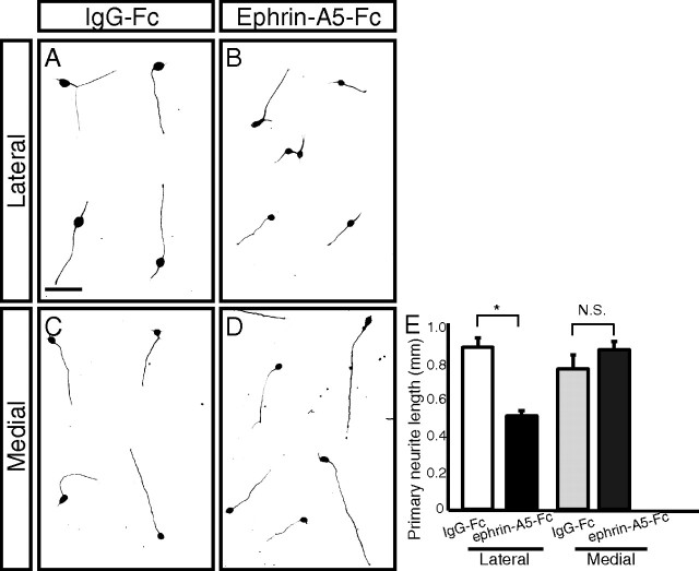
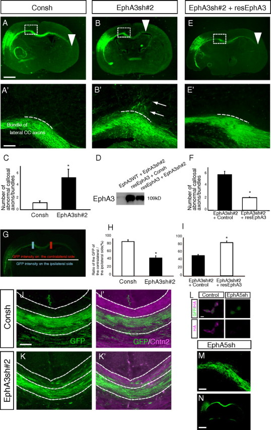
The corpus callosum, composed of callosal axons, is the largest structure among commissural connections in eutherian animals. Axon pathfinding of callosal neurons has been shown to be guided by intermediate targets, such as midline glial structures. However, it has not yet been understood completely how axon-axon interactions, another major mechanism for axon pathfinding, are involved in the pathfinding of callosal neurons. Here, we show that callosal axons from the medial and lateral regions of the mouse cerebral cortex pass through the dorsal and ventral parts, respectively, of the corpus callosum. Using an explant culture system, we observed that the axons from the medial and lateral cortices were segregated from each other in vitro, and that this segregation was attenuated by inhibition of EphA3 signaling. We also found that knockdown of EphA3, which is preferentially expressed in the lateral cortex, resulted in disorganized segregation of the callosal axons and disrupted axon pathfinding in vivo. These results together suggest the role of axonal segregation in the corpus callosum, mediated at least in part by EphA3, in correct pathfinding of callosal neurons.


Figures

Figure 1.
Figure 1.
DiI and DiO labeling of the medial and lateral cortices and the expression patterns of Cntn2 and Nrp1. A, Injection sites of DiI and DiO crystals. DiI and DiO were injected into the lateral and medial cortices, respectively. B, The labeled axons in a coronal section are shown at low magnification. C–E, The axons in the white box in B are shown at high magnification. The lateral cortical axons are segregated from the medial cortical axons in the CC. F, Injection sites of DiI and DiO crystals. DiO and DiI were injected into the medial-most (retrosplenial) cortex and a more lateral part of the medial cortex, respectively. G–I, The labeled axons in the CC are shown at high magnification. Both the axons of the retrosplenial cortex and the “more lateral” medial cortex passed through the dorsal part of the CC. J, K, In situ hybridization for Cntn2 and Nrp1. Yellow rectangles show the gradients of expression. L–Q, Immunohistochemistry for Cntn2 and Nrp1. The boxed regions in L–N are shown at high magnification (O–Q). Scale bars: B, J, L, 500 μm; C, G, O, 100 μm.
Figure 2.
Figure 2.
Interactions between axons originating from the medial (M) and lateral (L) cortical explants. A, Outline of the dissection method used in this experiment. B–D, The axons of the medial and lateral cortical explants were visualized by anti-GFP (green) and Tuj1 immunostaining (magenta). B′–D′, High magnification of the boxed regions in B–D. E–G, Other samples, which were processed in the same manner as described above. B/E, C/F, and D/G are cocultures of the medial and medial cortices, lateral and lateral cortices, and medial and lateral cortices, respectively. The medial and lateral cortical axons tended to be separated from each other, while the axons from homotypic explants were well mixed, suggesting the repulsive effect between the axons from medial and lateral cortical explants. Scale bars: B, E, 100 μm; B′, 25 μm.
Figure 3.
Figure 3.
Regionally preferential distribution of EphA3 and ephrin-A5. A, Schematic representation of the brain regions shown in B–E. B–E, In situ hybridization for EphA3 and ephrin-A5. EphA3 mRNA is expressed more strongly in the lateral cortex than in the medial cortex. On the other hand, ephrin-A5 mRNA is expressed preferentially in the medial cortex. EphA3 and ephrin-A5 mRNA are expressed in the presumptive layers II/III and layer V (Vanderhaeghen et al., 2000; Dufour et al., 2003). F–H, Protein localization of EphA3 and Cntn2. F′–H′, High magnification of F–H. Cntn2 protein is expressed specifically on the commissural axons that pass through the dorsal part of the CC, while EphA3 is detected on those in the ventral part. The expression of both molecules is mutually exclusive. I–K, Sagittal sections immunostained for EphA3 and Cntn2. HC, Hippocampal commissure. Scale bars: B, F, 500 μm; F′, 100 μm; I, 200 μm.
Figure 4.
Figure 4.
Ephrin-A5-Fc inhibits axonal growth of the lateral cortical neurons selectively. A–D, Primary dissociated culture of the lateral (A, B) or medial (C, D) cortices with the treatment of IgG-Fc or ephrin-A5-Fc. Ephrin-A5-Fc inhibited axonal growth of the lateral cortical neurons, while this inhibitory effect was not observed for the medial cortical neurons. E, Quantitative data of the effect of ephrin-A5-Fc on the medial and lateral cortical neurons. *p < 0.05. N.S., no significance. Scale bar: A, 50 μm.
Figure 5.
Figure 5.
Segregation of the medial and lateral cortical axons is attenuated by EphA3-Fc treatment and EphA3 knockdown in vitro. A–D, Cortical explants were prepared as described in Figure 2D. A, B, Coculture experiments with EphA3-Fc treatment. A, Cocultures of the medial and lateral cortices with added control IgG-Fc proteins. B, Cocultures of the medial and lateral cortices with EphA3-Fc treatment. A′ and B′ are high-magnification views of the boxed regions in A and B, respectively. The medial and lateral cortical axons, which were separated from each other in the in vitro control cultures (separated in 86%, 6 of 7 samples), were mixed with each other in the presence of EphA3-Fc (separated in 22%, 2 of 9 samples). C, Immunoblotting to examine the efficiency of knockdown by EphA3-silencing vectors on ectopically expressed EphA3. 293T cells were transfected with EphA3sh#1–#4 and control shRNA (Consh) vectors together with an EphA3 expression vector and the cell lysates were subjected to immunoblotting for EphA3. EphA3sh#2 (and also #3) knocked down EphA3 expression efficiently. D, E, Coculture experiments with the EphA3-knockdown lateral and the medial cortical axons. F, Schematic representation of the quantification method used in G. G, Quantification of the invasion rates of the lateral cortical axons into the medial cortical axons. *p < 0.05. Scale bars: A, 100 μm; A′, 50 μm; D, 200 μm.
Figure 6.
Figure 6.
Follower axon-specific introduction of plasmid vectors by in utero electroporation. A, B, Immunostaining for GFP (A) and TuJ1 (B) in the electroporation experiments. We transferred an empty pSilencer 3.0-H1 vector (Consh) together with the GFP vector on E12.5 and fixed the brains on E16.0. At E16.0, the most early follower axons had not yet passed through the CC (arrow in A), while pioneer axons from the cingulate cortex have already passed (arrowhead in B). C–F, Consh (C, D) or EphA3sh#2 (E, F) was transfected on E12.5, and the brains were fixed on E16.0. The tip of the growing axons of the EphA3-knockdown neurons reached almost the same point compared with that in the controls (yellow arrows in C, E). Scale bars: A, 500 μm; C, 200 μm.
Figure 7.
Figure 7.
EphA3 signaling is required for proper pathfinding of the callosal axons derived from the lateral cortex. A, B, E, Immunostaining for GFP in RNAi experiments for EphA3 (A, B) and the rescue experiments (E). A′, B′, and E′ are high-magnification images of the boxed regions in A, B, and E. Abnormal axons/bundles are indicated with white arrows in B′. The length of the longest axon was also decreased by EphA3-knockdown (white arrowheads in A and B). C, The number of abnormal axons/bundles in A′ and B′. We analyzed 23 brains (Consh, 11; EphA3sh#2, 12). D, Immunoblotting to examine the efficiency of rescue by resEphA3 for EphA3sh#2. 293T cells were transfected with the indicated plasmids, and the cell lysates were subjected to immunoblotting for EphA3. The resEphA3 plasmid appeared to be resistant to knockdown by EphA3sh#2. E, The elongation failure of callosal axons was rescued by cotransfection with the rescue vector (arrowhead in E). F, The number of abnormal axons/bundles in the rescue experiments. We analyzed 21 brains (EphA3sh#2+Control, 13; EphA3sh#2+resEphA3, 8). G, Schematic representation of the quantification method used in H and I. H, I, Quantitative data of the ratio of the intensity of GFP-positive axons leaving the CC to that of the GFP-positive axons entering the CC in the knockdown experiments (H) and the rescue experiments (I). J, K, Abnormal axons observed around the midline of the CC. The GFP-positive axons around the midline from the control (J) and EphA3-knockdown (K) experiments are shown. J′ and K′ show merged images of the GFP signals with immunostaining for Cntn2 (magenta). While most of the lateral cortical axons passed through the ventral part of the CC in the control, the EphA3-knockdown lateral cortical axons could not follow the normal axonal trajectory. L, Immunocytochemistry for HA to examine the efficiency of knockdown by EphA5-silencing vectors on ectopically expressed HA-EphA5. EphA5sh effectively knocked down the expression of EphA5. M, N, Immunostaining for GFP in RNAi experiment for EphA5. M, Normal axon pathfinding observed around the midline of the CC in the EphA5-knockdown neurons. N, Normal axonal elongation was also observed. *p < 0.05. Scale bars: A, N, 500 μm; A′, J, M, 100 μm; L, 10 μm.
Figure 8.
Figure 8.
Model of interaction between the medial and lateral callosal axons. The repulsive effects between the medial and lateral cortical axons contribute to the axonal segregation in the CC. Under normal conditions, lateral cortical axons would use the interaction with medial cortical axons to project to the contralateral neurons in the lateral cortex through the CC. When the functions of EphA3 are inhibited, the lateral cortical axons cannot interact with the medial cortical axons, disrupting their correct pathfinding and/or proper extension.

Similar articles

Cited by

References

    1. Bush JO, Soriano P. Ephrin-B1 regulates axon guidance by reverse signaling through a PDZ-dependent mechanism. Genes Dev. 2009;23:1586–1599. - PMC - PubMed
    1. Chan SO, Chung KY. Changes in axon arrangement in the retinofugal [correction of retinofungal] pathway of mouse embryos: confocal microscopy study using single- and double-dye label. J Comp Neurol. 1999;406:251–262. - PubMed
    1. Dufour A, Seibt J, Passante L, Depaepe V, Ciossek T, Frisén J, Kullander K, Flanagan JG, Polleux F, Vanderhaeghen P. Area specificity and topography of thalamocortical projections are controlled by ephrin/Eph genes. Neuron. 2003;39:453–465. - PubMed
    1. Egea J, Klein R. Bidirectional Eph-ephrin signaling during axon guidance. Trends Cell Biol. 2007;17:230–238. - PubMed
    1. Fame RM, MacDonald JL, Macklis JD. Development, specification, and diversity of callosal projection neurons. Trends Neurosci. 2011;34:41–50. - PMC - PubMed

Publication types

MeSH terms

LinkOut - more resources