

The Liprin homology domain is essential for the homomeric interaction of SYD-2/Liprin-α protein in presynaptic assembly
- PMID: 22072677
- PMCID: PMC3500560
- DOI: 10.1523/JNEUROSCI.0002-11.2011
The Liprin homology domain is essential for the homomeric interaction of SYD-2/Liprin-α protein in presynaptic assembly
Abstract
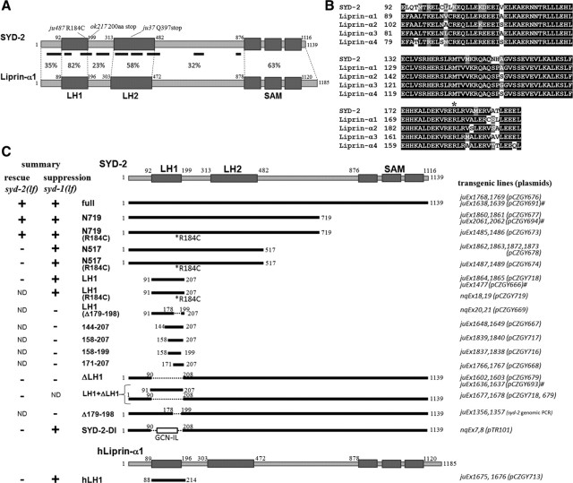
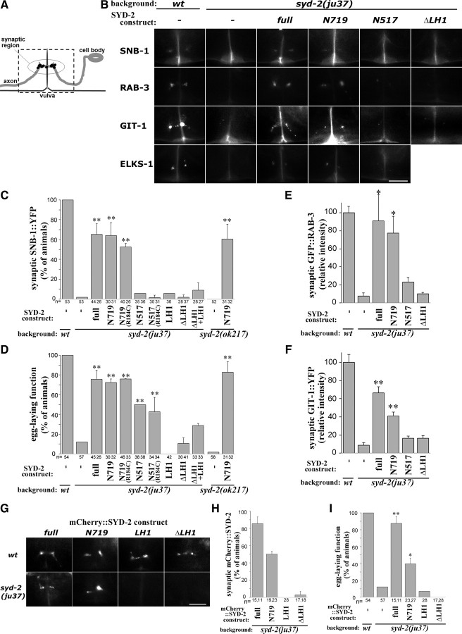
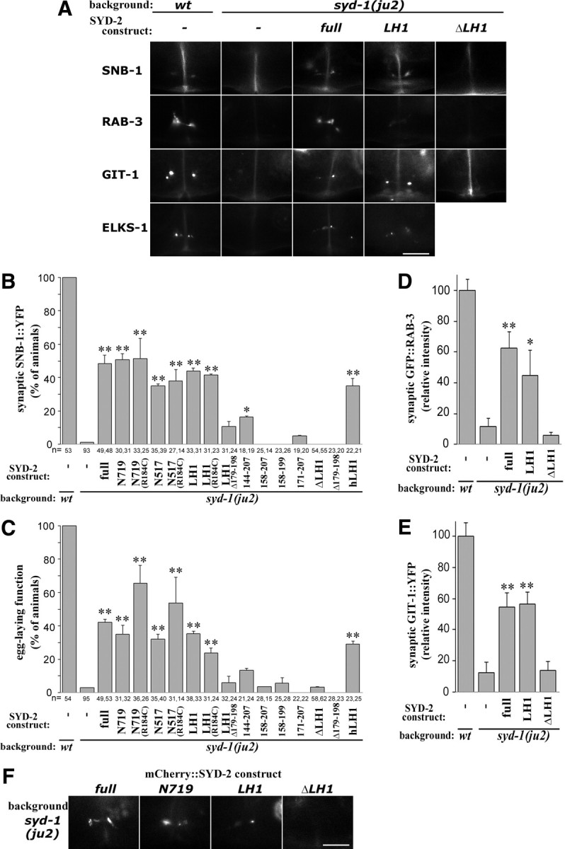
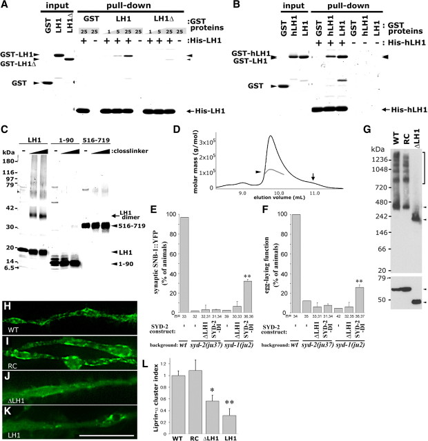
Synapses are asymmetric structures that are specialized for neuronal signal transduction. A unique set of proteins is present at the presynaptic active zone, which is a core structure essential for neurotransmitter release. In Caenorhabditis elegans HSN neurons, SYD-2, a Liprin-α family protein, acts together with a GAP protein SYD-1 to promote presynaptic assembly. Previous studies have shown that elevating the activity of syd-2 can bypass the requirement of syd-1. Liprin-α proteins are composed of coiled-coil-rich regions in the N-terminal half, which mediate interactions with adapter proteins at the presynaptic active zone, and three SAM domains in the C terminus, which bind proteins such as LAR receptor tyrosine phosphatase. To address the molecular mechanism by which SYD-2 activity is regulated, we performed structure-function studies. By monitoring the ability of SYD-2 transgenes to rescue syd-2(lf) and to suppress syd-1(lf) phenotypes in HSN neuron synapses, we identified the N-terminal half of SYD-2 as minimally required for rescuing syd-2(lf) phenotypes. A highly conserved short coiled-coil segment named Liprin Homology 1 (LH1) domain is both necessary and sufficient to suppress syd-1(lf) defects. We show that the LH1 domain forms a dimer and promotes further oligomerization and/or complex formation of SYD-2/Liprin-α proteins. The role of the LH1 domain in presynaptic assembly can be partially complemented by artificial dimerization. These findings suggest a model by which the self-assembly of SYD-2/Liprin-α proteins mediated by the coiled-coil LH1 domain is one of the key steps to the accumulation of presynaptic components at nascent synaptic junctions.
Figures
Similar articles
-
SYD-2 Liprin-alpha organizes presynaptic active zone formation through ELKS.Nat Neurosci. 2006 Dec;9(12):1479-87. doi: 10.1038/nn1808. Epub 2006 Nov 19. Nat Neurosci. 2006. PMID: 17115037
-
Intramolecular regulation of presynaptic scaffold protein SYD-2/liprin-α.Mol Cell Neurosci. 2013 Sep;56:76-84. doi: 10.1016/j.mcn.2013.03.004. Epub 2013 Mar 27. Mol Cell Neurosci. 2013. PMID: 23541703 Free PMC article.
-
Hierarchical assembly of presynaptic components in defined C. elegans synapses.Nat Neurosci. 2006 Dec;9(12):1488-98. doi: 10.1038/nn1806. Epub 2006 Nov 19. Nat Neurosci. 2006. PMID: 17115039 Free PMC article.
-
LAR, liprin alpha and the regulation of active zone morphogenesis.J Cell Sci. 2007 Nov 1;120(Pt 21):3723-8. doi: 10.1242/jcs.03491. J Cell Sci. 2007. PMID: 17959628 Review.
-
Liprin-α-Mediated Assemblies and Their Roles in Synapse Formation.Front Cell Dev Biol. 2021 Mar 19;9:653381. doi: 10.3389/fcell.2021.653381. eCollection 2021. Front Cell Dev Biol. 2021. PMID: 33869211 Free PMC article. Review.
Cited by
-
Submembrane liprin-α1 clusters spatially localize insulin granule fusion.J Cell Biol. 2025 Oct 6;224(10):e202410210. doi: 10.1083/jcb.202410210. Epub 2025 Aug 28. J Cell Biol. 2025. PMID: 40875978 Free PMC article.
-
The scaffolding protein SYD-2/Liprin-α regulates the mobility and polarized distribution of dense-core vesicles in C. elegans motor neurons.PLoS One. 2013;8(1):e54763. doi: 10.1371/journal.pone.0054763. Epub 2013 Jan 24. PLoS One. 2013. PMID: 23358451 Free PMC article.
-
RIMB-1/RIM-Binding Protein and UNC-10/RIM Redundantly Regulate Presynaptic Localization of the Voltage-Gated Calcium Channel in Caenorhabditis elegans.J Neurosci. 2019 Oct 30;39(44):8617-8631. doi: 10.1523/JNEUROSCI.0506-19.2019. Epub 2019 Sep 17. J Neurosci. 2019. PMID: 31530643 Free PMC article.
-
Structural basis of liprin-α-promoted LAR-RPTP clustering for modulation of phosphatase activity.Nat Commun. 2020 Jan 10;11(1):169. doi: 10.1038/s41467-019-13949-x. Nat Commun. 2020. PMID: 31924785 Free PMC article.
-
Liprin-α/SYD-2 determines the size of dense projections in presynaptic active zones in C. elegans.J Cell Biol. 2013 Dec 9;203(5):849-63. doi: 10.1083/jcb.201302022. J Cell Biol. 2013. PMID: 24322429 Free PMC article.
References
-
- Dai Y, Taru H, Deken SL, Grill B, Ackley B, Nonet ML, Jin Y. SYD-2 Liprin-alpha organizes presynaptic active zone formation through ELKS. Nat Neurosci. 2006;9:1479–1487. - PubMed
-
- Hallam SJ, Goncharov A, McEwen J, Baran R, Jin Y. SYD-1, a presynaptic protein with PDZ, C2 and rhoGAP-like domains, specifies axon identity in C. elegans. Nat Neurosci. 2002;5:1137–1146. - PubMed
Publication types
MeSH terms
Substances
Grants and funding
LinkOut - more resources
Full Text Sources
Molecular Biology Databases
Miscellaneous