. 2011 Nov 9;31(45):16279-91.
doi: 10.1523/JNEUROSCI.3835-11.2011.

Hippocampal GluA1-containing AMPA receptors mediate context-dependent sensitization to morphine

Affiliations

Hippocampal GluA1-containing AMPA receptors mediate context-dependent sensitization to morphine

Yan Xia et al. J Neurosci. .

Abstract

Glutamatergic systems, including AMPA receptors (AMPARs), are involved in opiate-induced neuronal and behavioral plasticity, although the mechanisms underlying these effects are not fully understood. In the present study, we investigated the effects of repeated morphine administration on AMPAR expression, synaptic plasticity, and context-dependent behavioral sensitization to morphine. We found that morphine treatment produced changes of synaptic AMPAR expression in the hippocampus, a brain area that is critically involved in learning and memory. These changes could be observed 1 week after the treatment, but only when mice developed context-dependent behavioral sensitization to morphine in which morphine treatment was associated with drug administration environment. Context-dependent behavioral sensitization to morphine was also associated with increased basal synaptic transmission and disrupted hippocampal long-term potentiation (LTP), whereas these effects were less robust when morphine administration was not paired with the drug administration environment. Interestingly, some effects may be related to the prior history of morphine exposure in the drug-associated environment, since alterations of AMPAR expression, basal synaptic transmission, and LTP were observed in mice that received a saline challenge 1 week after discontinuation of morphine treatment. Furthermore, we demonstrated that phosphorylation of GluA1 AMPAR subunit plays a critical role in the acquisition and expression of context-dependent behavioral sensitization, as this behavior is blocked by a viral vector that disrupts GluA1 phosphorylation. These data provide evidence that glutamatergic signaling in the hippocampus plays an important role in context-dependent sensitization to morphine and supports further investigation of glutamate-based strategies for treating opiate addiction.


Figures

Figure 1.
Figure 1.
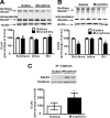
A–C, Administration of escalating doses of morphine in the home cage alters phosphorylation levels of GluA1 and GluA2 in the hippocampus. Twelve hours after treatment with escalating doses of morphine in the home cage, pGluA1 was increased in the homogenate, synaptosomal fraction, and the PSD relative to saline-treated mice, whereas pGluA2 was increased only in the total homogenate and PSD. Furthermore, repeated morphine treatment differentially alters synaptic targeting of GluA1 and GluA2 AMPAR subunits in the homogenate. D, The association between stargazin and GluA1 and the association between PSD95 and GluA1 were increased 12 h after treatment with escalating doses of morphine. E, The association between GRIP1 and GluA2 decreased 12 h after repeated morphine treatment, whereas no changes were observed in the association between PICK and GluA2. Inset, Representative blot showing stronger association of stargazin with GluA1 than with the GluA2 subunit. N = 3–4. *p < 0.05, morphine versus saline, unpaired t test. Error bars indicate SEM.
Figure 2.
Figure 2.
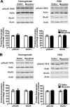
Administration of escalating doses of morphine in the home cage triggers internalization of the GluA2 subunit of AMPA receptors. A, B, Using a protein cross-linking assay with BS3, we found no changes in cell surface expression of GluA1 in the synaptosomal fraction, but GluA2 was decreased on the cell surface and in the surface/intracellular ratio 12 h after repeated morphine treatment. C, The association between clathrin and GluA2 was increased 12 h after repeated morphine treatment, suggesting that repeated morphine administration leads to an increased internalization of GluA2 subunits and thus decreases cell surface expression of GluA2. N = 3–4. *p < 0.05, morphine versus saline, unpaired t test. Error bars indicate SEM.
Figure 3.
Figure 3.
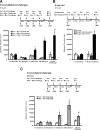
AMPAR expression was not altered 1 week after repeated morphine administration in the home cage. A, B, One week after the administration of escalating doses of morphine, no changes in GluA1 or GluA2 AMPAR subunits were observed in either the homogenate or the PSD, suggesting that alterations of AMPAR expression are not observed if morphine administration occurred in the home cage. Error bars indicate SEM.
Figure 4.
Figure 4.
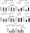
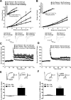
Behavioral sensitization following treatment with escalating doses of morphine is selectively expressed in a drug-associated environment. A–C, A schematic of the design is shown for either context-dependent behavioral sensitization or the unpaired group. LMA indicates that locomotor activity was recorded immediately following injection. A, The effects of escalating doses of morphine on context-dependent behavioral sensitization. Mice treated with escalating doses of morphine exhibited increased context-dependent locomotor activity when compared with saline-treated mice and with mice that only received 5 mg/kg morphine on the challenge day (*). Furthermore, mice treated with escalating doses of morphine had increased locomotor activity during the 1 week challenge of 5 mg/kg morphine when compared with the initial effects of 5 mg/kg morphine on locomotor activity (#). B, In contrast, behavioral sensitization to the effects of 5 mg/kg morphine was not observed when morphine administration was not consistently paired with the locomotor activity chambers (morphine unpaired). C, Mice treated with a saline challenge after context-dependent morphine treatment exhibited a conditioned response when compared with saline-treated mice (*) and had increased locomotor activity after the 1 week saline challenge when compared with the initial effects of the first 5 mg/kg injection of morphine (#). N = 8. *,#p < 0.05, one-way ANOVA with Tukey's post hoc comparisons or unpaired t tests. Error bars indicate SEM.
Figure 5.
Figure 5.
Alterations in synaptic distribution of AMPARs following context-dependent behavioral sensitization to morphine. A, Hippocampal tissue from saline- and morphine-treated mice was collected after context-dependent behavioral sensitization followed by a morphine challenge 7 d later (paired group). Context-dependent treatment of morphine produced an increase in pGluA1 and GluA1 in the total homogenate, whereas no effects were observed in pGluA2 and GluA2 levels. Furthermore, context-dependent treatment of morphine produced an increase in pGluA1 and decreased GluA2 levels in the PSD. B, Hippocampal tissue from saline- and morphine-treated mice was collected after context-dependent behavioral sensitization followed by a saline challenge 7 d later (paired group). Mice that received a saline challenge 1 week after context-dependent morphine treatment showed increased GluA1 and decreased pGluA2 in the PSD, whereas pGluA1 was decreased in the total homogenate. C, In contrast, no changes in synaptic GluA1 or GluA2 were observed when mice were treated with escalating doses of morphine in an unpaired design, but pGluA2 levels were increased in the homogenate. S+S, Saline plus Saline Challenge; S+M, Saline plus Morphine Challenge; M+M, Morphine plus Morphine Challenge; M+S, Morphine plus Saline Challenge. N = 3–4. *p < 0.05, one-way ANOVA with Tukey's post hoc comparisons or unpaired t test. Error bars indicate SEM.
Figure 6.
Figure 6.
Increased synaptic transmission and disrupted LTP following context-dependent behavioral sensitization to morphine. A, Mice treated with morphine in a paired design that received a morphine challenge showed a significantly greater fEPSP slope relative to saline-treated mice (*). Mice that were administered morphine in an unpaired design exhibited an intermediate stimulus–response slope that was significantly different from both morphine-paired mice (#) and saline-treated mice (*). B, Mice treated with morphine in a paired design that received a saline challenge exhibited a significantly enhanced fEPSP slope compared with saline-treated mice (*). The insets show fEPSP–FV relationships. C, Mice that received a morphine challenge 7 d after context-dependent morphine treatment showed disrupted LTP when compared with saline-treated mice, whereas mice that received unpaired morphine showed an intermediate effect relative to morphine-paired and saline-treated mice. D, Mice that received a saline challenge 7 d after context-dependent morphine treatment showed impaired LTP when compared with saline-treated mice. E, F, The histograms show an average percentage inhibition (mean ± SEM) of EPSCs recorded from hippocampal slices. Sample traces (averages of 20 trials) were recorded before and after naspm application (100 μm, 5 min). Naspm reduced the amplitude of EPSCs in mice that received context-dependent morphine treatment (*), regardless of whether a saline or morphine challenge was administered 1 week later. N = 4–6. *,#p < 0.05, one-way ANOVA with Tukey's post hoc comparisons or unpaired t tests. Error bars indicate SEM.
Figure 7.
Figure 7.
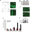
GluA1 S845 phosphorylation in the hippocampus is required for context-dependent behavioral sensitization to morphine. A, Representative examples of HSV infection in the hippocampus. Mice received hippocampal infusions of HSV-GFP, and tissue was collected 3 d after infection. The area of infection is indicated by HSV-GFP-labeled neurons, and diagrams from a mouse brain atlas (Franklin and Paxinos, 2007) indicate the hippocampal regions targeted for viral infusion. B, Infusions of HSV-S845A into the hippocampus decreased S845 GluA1 phosphorylation (3 d after infection) in naive mice but did not alter total levels of GluA1. C, Representative hippocampal slices stained with TUNEL, a marker of DNA fragmentation. Overexpression of S845A did not produce any observable changes in neurotoxicity in the hippocampus 5 d after HSV-S845A infusion. D, Mice treated with escalating doses of morphine that received a HSV-lacZ control protein exhibited increased context-dependent locomotor activity when compared with all other groups, whereas morphine-treated mice that received hippocampal infusions of HSV-S845A did not exhibit context-dependent behavioral sensitization to morphine. E, Infusion of HSV-S845A disrupted the association between GluA1 and stargazin that is observed following context-dependent sensitization to morphine. N = 6–7. *p < 0.05, one-way ANOVA with Tukey's post hoc comparisons or unpaired t tests. Error bars indicate SEM.

Similar articles

Cited by

References

    1. Barrot M, Olivier JD, Perrotti LI, DiLeone RJ, Berton O, Eisch AJ, Impey S, Storm DR, Neve RL, Yin JC, Zachariou V, Nestler EJ. CREB activity in the nucleus accumbens shell controls gating of behavioral responses to emotional stimuli. Proc Natl Acad Sci U S A. 2002;99:11435–11440. - PMC - PubMed
    1. Billa SK, Sinha N, Rudrabhatla SR, Morón JA. Extinction of morphine-dependent conditioned behavior is associated with increased phosphorylation of the GluR1 subunit of AMPA receptors at hippocampal synapses. Eur J Neurosci. 2009;29:55–64. - PMC - PubMed
    1. Billa SK, Liu J, Bjorklund NL, Sinha N, Fu Y, Shinnick-Gallagher P, Morón JA. Increased insertion of glutamate receptor 2-lacking alpha-amino-3-hydroxy-5-methyl-4-isoxazole propionic acid (AMPA) receptors at hippocampal synapses upon repeated morphine administration. Mol Pharmacol. 2010;77:874–883. - PMC - PubMed
    1. Borgkvist A, Valjent E, Santini E, Hervé D, Girault JA, Fisone G. Delayed, context- and dopamine D1 receptor-dependent activation of ERK in morphine-sensitized mice. Neuropharmacology. 2008;55:230–237. - PubMed
    1. Bossert JM, Stern AL, Theberge FR, Cifani C, Koya E, Hope BT, Shaham Y. Ventral medial prefrontal cortex neuronal ensembles mediate context-induced relapse to heroin. Nat Neurosci. 2011;14:420–422. - PMC - PubMed

Publication types

MeSH terms

LinkOut - more resources