. 2011 Nov 9;31(45):16410-22.
doi: 10.1523/JNEUROSCI.3280-11.2011.

Phox2b-expressing neurons of the parafacial region regulate breathing rate, inspiration, and expiration in conscious rats

Affiliations

Phox2b-expressing neurons of the parafacial region regulate breathing rate, inspiration, and expiration in conscious rats

Stephen B G Abbott et al. J Neurosci. .

Abstract

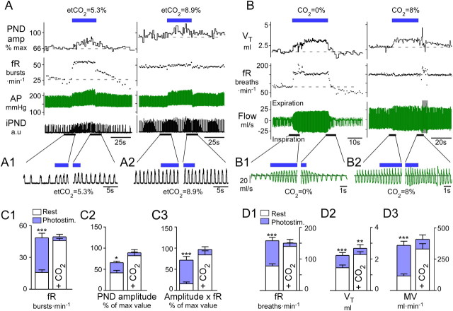
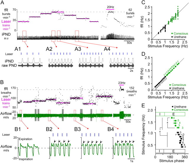
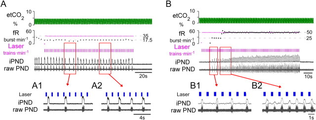
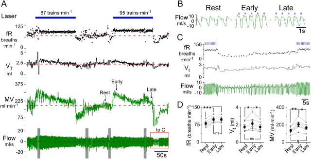
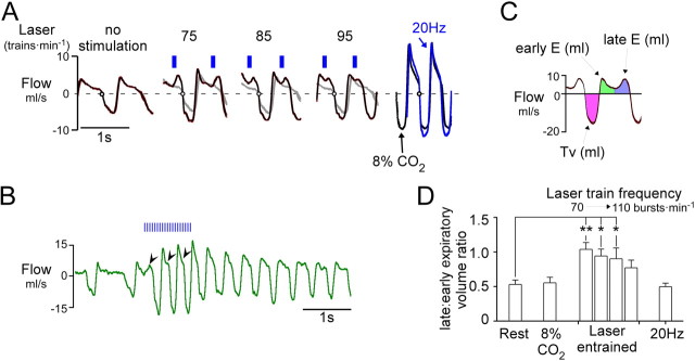
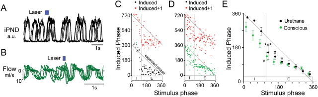
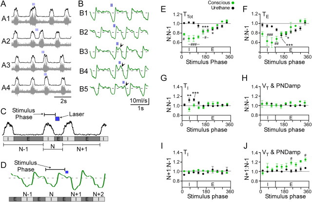
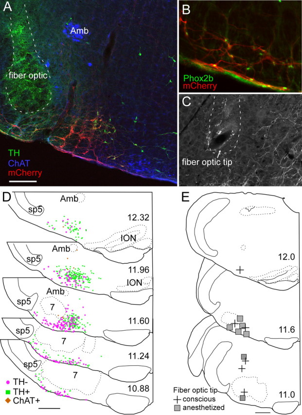
The retrotrapezoid nucleus contains Phox2b-expressing glutamatergic neurons (RTN-Phox2b neurons) that regulate breathing in a CO₂-dependent manner. Here we use channelrhodopsin-based optogenetics to explore how these neurons control breathing in conscious and anesthetized adult rats. Respiratory entrainment (pacing) of breathing frequency (fR) was produced over 57% (anesthetized) and 28% (conscious) of the natural frequency range by burst activation of RTN-Phox2b neurons (3-8 × 0.5-20 ms pulses at 20 Hz). In conscious rats, pacing under normocapnic conditions increased tidal volume (V(T)) and each inspiration was preceded by active expiration, denoting abdominal muscle contraction. During long-term pacing V(T) returned to prestimulation levels, suggesting that central chemoreceptors such as RTN-Phox2b neurons regulate V(T) partly independently of their effect on fR. Randomly applied light trains reset the respiratory rhythm and shortened the expiratory phase when the stimulus coincided with late-inspiration or early-expiration. Importantly, continuous (20 Hz) photostimulation of the RTN-Phox2b neurons and a saturating CO₂ concentration produced similar effects on breathing that were much larger than those elicited by phasic RTN stimulation. In sum, consistent with their anatomical projections, RTN-Phox2b neurons regulate lung ventilation by controlling breathing frequency, inspiration, and active expiration. Adult RTN-Phox2b neurons can entrain the respiratory rhythm if their discharge is artificially synchronized, but continuous activation of these neurons is much more effective at increasing lung ventilation. These results suggest that RTN-Phox2b neurons are no longer rhythmogenic in adulthood and that their average discharge rate may be far more important than their discharge pattern in driving lung ventilation.


Figures

Figure 1.
Figure 1.
Breathing stimulation elicited by photostimulation of RTN Phox2b neurons is occluded by hypercapnia. A, Urethane-anesthetized, vagotomized, and ventilated rat. Left, Normocapnia (etCO2, 5.3%). Right, Hypercapnia (etCO2, 8.9%). RTN photostimulation (20 Hz, 5 ms pulses) is indicated by the blue bar. Under normocapnia, respiratory frequency, fR, increased within seconds to a steady-state level >50 bursts · min−1 and decayed in a two-step fashion (rapid, then slow) upon cessation of the stimulus. Phrenic nerve discharge peak amplitude, PNDamp, progressively increased during the stimulus and also had a slow decay upon cessation of the stimulus. During hyperoxic hypercapnia (right) the same stimulus produced no further increase of fR, a lesser increase in peak amplitude, but an identical increase in arterial pressure (AP). B, Conscious rat in flow-through, whole-body plethysmography chamber. Left, Hyperoxic normocapnia (100% O2); right, hypercapnia (8% CO2, balance O2). Continuous RTN stimulation (23 Hz, 3 ms pulses, 30 s total duration, blue bar) raised breathing frequency, fR, and tidal volume, VT, with identical onset kinetics as that in the anesthetized rat. Following the end of the stimulus, fR and VT dropped precipitously below baseline in the conscious rat, presumably because of hyperventilation-induced hypocapnia. During hyperoxic hypercapnia (right), RTN photostimulation produced a small increase in VT but no increase in fR. A1, A2, B1, B2, Expanded traces highlighting the periods surrounding the beginning and end of the stimulus. Note that in plethysmography records, inspiration is downward. C, Group data for anesthetized rats. From left to right: C1, fR; C2, PND peak amplitude; and C3, double product (fR × PND amplitude) at normal (left columns) and elevated CO2 (right columns). D, Group data for conscious rats. From left to right: D1, fR; D2, VT; and D3, double product MV, minute volume, at normal (left columns) and elevated CO2 (right, 8% CO2 in pure oxygen). *** p < 0.001, **p < 0.01, *p < 0.05 between prestimulus baseline and peak values during the stimulation period; two-way ANOVA with multiple comparisons.
Figure 2.
Figure 2.
Respiratory pacing by intermittent photostimulation of RTN neurons in anesthetized and conscious rats. A, Urethane-anesthetized rat. Photostimulus trains (4 × 20 ms pulses at 20 Hz) entrained PND over a range of 32 breaths · min−1 (from 25 to 60 bursts · min−1; see also excerpts A1, A2). The maximal fR observed in this rat during continuous RTN stimulation at 20 Hz was 62 bursts · min−1 (at right in A). At low frequencies (A1) the laser trains settled in late-expiration, whereas at higher frequencies the stimulus train settled earlier during the expiratory phase (A2). RTN stimulation at 60 trains · min−1 caused arrhythmia (excerpt A3). A4 shows that fR follows the transition from one stimulus frequency to the next (50 to 40 trains · min−1). B, Conscious quiet rat. Light pulses (3 × 3 ms pulses at 20 Hz) entrained fR in the range of 75–120 breaths · min−1. Resting fR was close to 60 min−1 in this rat, and the maximum fR seen in presence of high CO2 was 152 breaths · min−1. Continuous high-frequency stimulation (23 Hz, 3 ms, at right in B) produced a more robust activation of breathing than short bursts. Gray bars indicate periods of behavioral artifacts (e.g., sniffing, movement). B1B4 excerpts from B show expanded plethysmography traces at various entrainment frequencies. B1, Quiet breathing in the absence of stimulation. Note monophasic decay of airflow during expiration consistent with passive lung recoil. B2B4, Airflow traces at three different pacing frequencies. Expiratory flow becomes biphasic with pronounced increase in flow during late-expiration (arrowheads), indicative of active expiration. Note that the stimulation trains settle in mid-expiration during entrainment. C, Relationship between stimulus train frequency and fR for the rats shown in A (anesthetized, black) or B (conscious, green). The instantaneous frequency of each burst/breath is plotted against the frequency of the light trains. Note the increased variability of fR at higher entrainment frequencies, reflecting poor entrainment. D, Group data showing the reliability of entrainment at a range of stimulus frequencies in anesthetized (N = 7) and conscious (N = 6) animals. Each point represents the average fR (±SEM) when entrainment was successful. Adjacent numbers indicate the number of animals that were successfully entrained at a given stimulation frequency. Not every frequency was tested in every animal; hence, the varying number of determinations for each tested frequency. E, Relationship between the stimulus train frequency and the phase angle of the stimulus train during successful entrainment (E) of the breathing cycle. Note that as stimulus (and entrainment) frequency increased, the phase angle of the stimulus train decreased. In conscious animals, the stimulus occurred at a significantly earlier phase in the respiratory cycle than in anesthetized rats. (*p < 0.05).
Figure 3.
Figure 3.
RTN photostimulation produces quantal breathing in urethane–anesthetized hypocapnic rats. A, When ventilation was adjusted to maintained etCO2 close to the apneic threshold trains of laser light (7 × 5 ms pulses at 20 Hz; train frequency, 35 min−1) entrained PND in a 3:1 (A1), or 2:1 ratio (A2). B, Quantal entrainment in a different anesthetized rat with etCO2 maintained below apneic threshold. Laser trains (7 × 5 ms pulse at 20 Hz; train frequency, 50 min−1) delivered during phrenic apnea first produced quantal entrainment in a 2:1 ratio (B1), eventually achieving 1:1 entrainment (B2).
Figure 4.
Figure 4.
Respiratory pacing above resting frequency causes compensatory decreases in tidal volume in conscious rats. A, Conscious quiet rat breathing pure oxygen. Prolonged respiratory pacing (3 × 5 ms pulses at 20 Hz; train frequency, 87 and 95 trains · min−1) caused a gradual reduction of the tidal volume, VT, to the resting (prestimulation) level, and a parallel decline in total ventilation, MV. After the end of the stimulus, tidal volume and frequency were transiently reduced, reflecting the hypocapnia caused by an incomplete return of MV to prestimulation levels. Gray bars in flow trace indicate behavioral disturbances. B, Expanded traces from A showing the compensatory changes in VT during long periods of entrainment (147 s at 95 trains · min−1). During the early part of the stimulation (middle, “Early”), VT was increased compared to the resting state (left). Two and a half minutes later (right, “Late”), VT had decreased to prestimulation levels. C, Expanded trace from A (red box) illustrating the after stimulus hypoventilation that followed 147 s of pacing at the rate of 95 trains min−1. fR fell to 28 breaths · min−1 before returning to resting levels. D, Group data (N = 6) summarizing frequency of respiration (fR), tidal volume (VT) and total ventilation (MV) at rest vs during the early and late part of the entrainment period (***p < 0.001, **p < 0.01, *p < 0.05; one-way ANOVA).
Figure 5.
Figure 5.
RTN photostimulation increases late-expiratory flow in conscious rats. A, Trajectory of respiratory flow (waveform average, 10–15 breaths) at rest and during respiratory pacing caused by burst photostimulation of RTN at various frequencies (no stimulation, 75, 85, and 95 min−1). Traces at extreme right depict flow trajectory during continuous 20 Hz stimulus (blue trace) or during hypercapnia (8% CO2; black trace) in the same rat. At rest (left excerpt) during quiet breathing (72 breaths · min−1) expiratory flow was monotonically decremental, reflecting the passive recoil of the lungs and no active expiration. When breathing was entrained at 75, 85, and 95 breaths · min−1 respectively (middle), a large positive peak in flow preceded inspiration. This peak was most prominent at lower frequencies of entrainment and tended to fuse with the early-expiratory peak at higher frequencies. During high-frequency continuous stimulation (20 Hz; right, blue) under hypercapnic conditions (8% inspired; right, black), the expiratory flow became monophasic, although active expiration must have been present. B, Original record showing the effect of a 1 s stimulus train (20 Hz, 3 ms pulse). This example illustrates the progressive change in the pattern of expiratory flow from a biphasic to a monophasic pattern during continuous high-frequency photostimulation. C, Tidal volume, early and late-expiratory volumes are color coded; early and late volumes were defined by dividing expiration (E) in two periods of equal duration. D, Group data (N = 6). During stimulation-induced respiratory pacing, the ratio between late-expiratory and early-expiratory volume was significantly increased relative to resting breathing (**p < 0.01, *p < 0.05 relative to rest; one-way ANOVA). The imbalance between late-expiratory and early-expiratory flow was most prominent at the lowest entrainment frequencies. When respiratory frequency was elevated by hypercapnia or continuous RTN stimulation at 20 Hz, the ratio between late-expiratory and early-expiratory volume was no different from that observed at rest.
Figure 6.
Figure 6.
Low-frequency train stimulation resets the respiratory cycle. A, Overlaid recordings of integrated PND from one anesthetized rat. The waveforms are taken from periods during low-frequency (<0.2 Hz) train stimulation and are aligned to the onset of the stimulus (six traces in total). When the stimulus was delivered during expiration (in all cases), an advanced inspiratory burst occurred and the respiratory rhythm was reset as shown by the synchronization of PND activity following stimulation. B, Overlaid flow traces from one conscious rat with description as per A. C, D, Phase resetting curves from a single anesthetized (C) and conscious rat (D). In both cases, a stimulus delivered during expiration dramatically reduced the induced phase (stimulus onset to following inspiration) and reset the respiratory rhythm (i.e., N + 1 induced phase ≈ 360°). E, Grouped data from anesthetized (N = 4) and conscious experiments (N = 6) showing that stimulus-induced phase resetting occurs preferentially during early-expiration in anesthetized rats and conscious rats. Stimulation during late-inspiration did not significantly reset the respiratory rhythm in anesthetized animals or conscious rats (***p < 0.001, *p < 0.05 vs N + 1 cycle in anesthetized rats; #p < 0.05 vs N + 1 cycle in conscious rats two-way ANOVA).
Figure 7.
Figure 7.
Phase-dependent perturbations of respiratory frequency and amplitude in anesthetized and conscious rats. RTN photostimulation was applied at frequencies less than the resting breathing frequency (<0.2 Hz) so that the stimulus occurred at random within the breathing cycle. A, Original recordings of integrated (top traces) and raw PND (bottom traces) in an anesthetized rat. Train stimulation produced phase-specific perturbations in total respiratory cycle (TTot), inspiratory time (TI), expiratory time (TE), and peak PND amplitude (PNDamp). In this example, stimulation during late-inspiration (A1) increased PNDamp and TI. When trains occurred during expiration (A2, A3) there was a decrease in TE with a modest increase in PNDamp, but no change in TI. When the stimulus coincided with the spontaneous phrenic burst, PNDamp was modestly increased and there was no change in respiratory timing (A4). B, Respiratory flow in a conscious rat. As in A, brief trains (3 pulses at 20 Hz) produced phase-specific perturbations in TTot, TI, TE, and tidal volume, VT. Stimulation during early-inspiration increased VT (B1). Stimulation during late-inspiration reduced the expiratory phase (B2). Stimulation during early- to mid-expiration decreased TE and produced active expiration (B3, B4). Stimulation during late-expiration (B5) shortened TE modestly and increased VT of the next inspiration. C, D, Data analysis from experiments in anesthetized (C) and conscious (D) rats. See Materials and Methods for more details. EJ, Group data from anesthetized (N = 4) and conscious experiments (N = 5). E, Effect of stimulus on respiratory period (TTot). F, Effect of stimulus on expiratory duration (TE). G, Effect of stimulus on inspiratory duration (TI). H, Effect of stimulus on tidal volume or PNDamp. In EH, values are expressed as ratio between respiratory cycles N and N − 1 (see definitions in C and D). A value of 1 indicates no change from the N − 1 (control) cycle. Note that in both conditions we did not observe significant increases in TE. I, J, Group data from anesthetized (N = 4) and conscious experiments (N = 5) showing the changes in PNDamp or VT in the N + 1 cycle relative to the control cycle N − 1. Brief stimulation during late-expiration significantly increased VT but not PNDamp of the following inspiratory effort (N + 1 cycle) (***p < 0.001, *p < 0.05 vs N + 1 for EH and N + 2 for I, J in anesthetized rats; ###p < 0.001, ##p < 0.01, #p < 0.05 vs N + 1 for EH and N + 2 for I, J in conscious rats).
Figure 8.
Figure 8.
Anatomical distribution of ChR2-transfected cells and fiber optic locations. A, Example of fiber optic location (dotted line, the green color within the fiber optic tract is an auto fluorescence artifact) next to the ChR2-mCherry-transfected neurons (mCherry in red). Choline acetyl-transferase-immunoreactive neurons (represented in blue) located in the ambiguus nucleus (Amb) and medial to the retrotrapezoid nucleus are not transfected. Several mCherry-positive neurons in this transverse section are catecholaminergic (tyrosine hydroxylase, TH, in green). These neurons generally occupy a medial location relative to the noncatecholaminergic, noncholinergic neurons (the RTN-Phox2b neurons). B, Native mCherry fluorescence (red) detected along with Phox2b-immunoreactivity (green nuclei). Note the presence of Phox2b in every transfected neuron, including within the marginal layer of the retrotrapezoid nucleus. C, Example of a fiber optic tract (dotted line) found to end next to the labeled axons of ChR2-mCherry-transfected neurons. D, Caudal-to-rostral series of transverse sections (bregma levels in millimeters as indicated) depicting the location of all the ChR2-mCherry-transfected neurons identified in 12 cases. Computer-generated drawings with each ChR2-mCherry-transfected neuron indicated by either a green square (TH+, TH-immunoreactive), brown diamond (ChAT+, choline acetyl transferase-immunoreactive), or magenta circle (TH-, neither TH-immunoreactive nor ChAT-immunoreactive) from the respective sections indicated for each bregma level from 12 animals were optimally superimposed using landmarks such as the ventral medullary surface, the edge of the trigeminal tract, the compact portion of nucleus ambiguus, the inferior olive, and the facial motor nucleus. E, Location of fiber optic tips plotted on three transverse sections at the indicated bregma level (in mm). Scale bars: (in A) A, C, 200 μm, B, 100 μm; (in D, E), 500 μm.

References

    1. Abbott SB, Stornetta RL, Fortuna MG, Depuy SD, West GH, Harris TE, Guyenet PG. Photostimulation of retrotrapezoid nucleus Phox2b-expressing neurons in vivo produces long-lasting activation of breathing in rats. J Neurosci. 2009a;29:5806–5819. - PMC - PubMed
    1. Abbott SB, Stornetta RL, Socolovsky CS, West GH, Guyenet PG. Photostimulation of channelrhodopsin-2 expressing ventrolateral medullary neurons increases sympathetic nerve activity and blood pressure in rats. J Physiol. 2009b;587:5613–5631. - PMC - PubMed
    1. Abdala AP, Rybak IA, Smith JC, Paton JF. Abdominal expiratory activity in the rat brainstem-spinal cord in situ: patterns, origins, and implications for respiratory rhythm generation. J Physiol. 2009a;587:3539–3559. - PMC - PubMed
    1. Abdala AP, Rybak IA, Smith JC, Zoccal DB, Machado BH, St-John WM, Paton JF. Multiple pontomedullary mechanisms of respiratory rhythmogenesis. Respir Physiol Neurobiol. 2009b;168:19–25. - PMC - PubMed
    1. Ballanyi K, Ruangkittisakul A, Onimaru H. Opioids prolong and anoxia shortens delay between onset of preinspiratory (pFRG) and inspiratory (preBotC) network bursting in newborn rat brainstems. Pflugers Arch. 2009;458:571–587. - PubMed

Publication types

MeSH terms

LinkOut - more resources