The anticonvulsant response to valproate in kindled rats is correlated with its effect on neuronal firing in the substantia nigra pars reticulata: a new mechanism of pharmacoresistance
- PMID: 22072692
- PMCID: PMC6633222
- DOI: 10.1523/JNEUROSCI.2506-11.2011
The anticonvulsant response to valproate in kindled rats is correlated with its effect on neuronal firing in the substantia nigra pars reticulata: a new mechanism of pharmacoresistance
Abstract
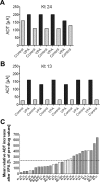
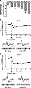
Resistance to antiepileptic drugs (AEDs) is a major problem in epilepsy treatment. However, mechanisms of resistance are only incompletely understood. We have recently shown that repeated administration of the AED phenytoin allows selecting resistant and responsive rats from the amygdala kindling model of epilepsy, providing a tool to study mechanisms of AED resistance. We now tested whether individual amygdala-kindled rats also differ in their anticonvulsant response to the major AED valproate (VPA) and which mechanism may underlie the different response to VPA. VPA has been proposed to act, at least in part, by reducing spontaneous activity in the substantia nigra pars reticulata (SNr), a main basal ganglia output structure involved in seizure propagation, seizure control, and epilepsy-induced neuroplasticity. Thus, we evaluated whether poor anticonvulsant response to VPA is correlated with low efficacy of VPA on SNr firing rate and pattern in kindled rats. We found (1) that good and poor VPA responders can be selected in kindled rats by repeatedly determining the effect of VPA on the electrographic seizure threshold, and (2) a significant correlation between the anticonvulsant response to VPA in kindled rats and its effect on SNr firing rate and pattern. The less VPA was able to raise seizure threshold, the lower was the VPA-induced reduction of SNr firing rate and the VPA-induced regularity of SNr firing. The data demonstrate for the first time an involvement of the SNr in pharmacoresistant experimental epilepsy and emphasize the relevance of the basal ganglia as target structures for new treatment options.
Figures


Similar articles
-
Subregional changes in discharge rate, pattern, and drug sensitivity of putative GABAergic nigral neurons in the kindling model of epilepsy.Eur J Neurosci. 2004 Nov;20(9):2377-86. doi: 10.1111/j.1460-9568.2004.03699.x. Eur J Neurosci. 2004. PMID: 15525279
-
A comparison of the effects of valproate and its major active metabolite E-2-en-valproate on single unit activity of substantia nigra pars reticulata neurons in rats.J Pharmacol Exp Ther. 1996 Jun;277(3):1305-14. J Pharmacol Exp Ther. 1996. PMID: 8667191
-
Reduction in firing rate of substantia nigra pars reticulata neurons by valproate: influence of different types of anesthesia in rats.Brain Res. 1995 Dec 8;702(1-2):133-44. doi: 10.1016/0006-8993(95)01030-4. Brain Res. 1995. PMID: 8846068
-
Animal models of drug-resistant epilepsy.Novartis Found Symp. 2002;243:149-59; discussion 159-66, 180-5. Novartis Found Symp. 2002. PMID: 11990774 Review.
-
A speculative model of affective illness cyclicity based on patterns of drug tolerance observed in amygdala-kindled seizures.Mol Neurobiol. 1996 Aug;13(1):33-60. doi: 10.1007/BF02740751. Mol Neurobiol. 1996. PMID: 8892335 Review.
Cited by
-
Not Part of the Temporal Lobe, but Still of Importance? Substantia Nigra and Subthalamic Nucleus in Epilepsy.Front Syst Neurosci. 2020 Dec 2;14:581826. doi: 10.3389/fnsys.2020.581826. eCollection 2020. Front Syst Neurosci. 2020. PMID: 33381016 Free PMC article. Review.
-
Empagliflozin Mitigates PTZ-Induced Seizures in Rats: Modulating Npas4 and CREB-BDNF Signaling Pathway.J Neuroimmune Pharmacol. 2025 Jan 7;20(1):5. doi: 10.1007/s11481-024-10162-6. J Neuroimmune Pharmacol. 2025. PMID: 39776284 Free PMC article.
-
Drug Resistance in Epilepsy: Clinical Impact, Potential Mechanisms, and New Innovative Treatment Options.Pharmacol Rev. 2020 Jul;72(3):606-638. doi: 10.1124/pr.120.019539. Pharmacol Rev. 2020. PMID: 32540959 Free PMC article. Review.
-
Translational Considerations in the Development of Intranasal Treatments for Epilepsy.Pharmaceutics. 2023 Jan 10;15(1):233. doi: 10.3390/pharmaceutics15010233. Pharmaceutics. 2023. PMID: 36678862 Free PMC article. Review.
-
Animal Models of Drug-Resistant Epilepsy as Tools for Deciphering the Cellular and Molecular Mechanisms of Pharmacoresistance and Discovering More Effective Treatments.Cells. 2023 Apr 24;12(9):1233. doi: 10.3390/cells12091233. Cells. 2023. PMID: 37174633 Free PMC article. Review.
References
-
- Albala BJ, Moshé SL, Okada R. Kainic-acid-induced seizures: a developmental study. Brain Res. 1984;315:139–148. - PubMed
-
- Deniau JM, Hammond C, Riszk A, Feger J. Electrophysiological properties of identified output neurons of the rat substantia nigra (pars compacta and pars reticulata): evidences for the existence of branched neurons. Exp Brain Res. 1978;32:409–422. - PubMed
-
- Depaulis A, Vergnes M, Marescaux C. Endogenous control of epilepsy: the nigral inhibitory system. Prog Neurobiol. 1994;42:33–52. - PubMed
-
- De Sarro G, Meldrum BS, Reavill C. Anticonvulsant action of 2-amino-7-phosphonoheptanoic acid in the substantia nigra. Eur J Pharmacol. 1984;106:175–179. - PubMed
-
- De Sarro G, Patel S, Meldrum BS. Anticonvulsant action of a kainate antagonist gamma-d-glutamyl aminomethylsulphonic acid injected focally into the substantia nigra and entopeduncular nucleus. Eur J Pharmacol. 1986;132:229–236. - PubMed
Publication types
MeSH terms
Substances
LinkOut - more resources
Full Text Sources
Miscellaneous
