

UHRF1 phosphorylation by cyclin A2/cyclin-dependent kinase 2 is required for zebrafish embryogenesis
- PMID: 22072796
- PMCID: PMC3248904
- DOI: 10.1091/mbc.E11-06-0487
UHRF1 phosphorylation by cyclin A2/cyclin-dependent kinase 2 is required for zebrafish embryogenesis
Abstract
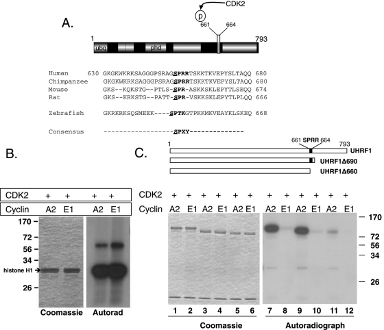
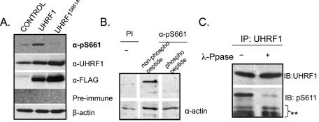
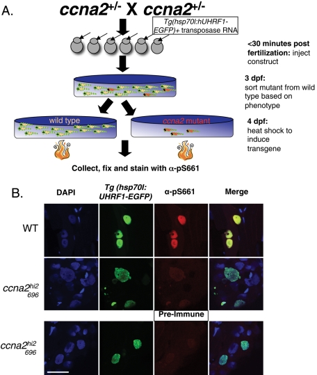
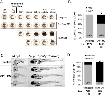
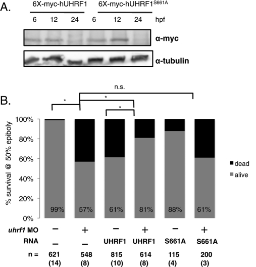
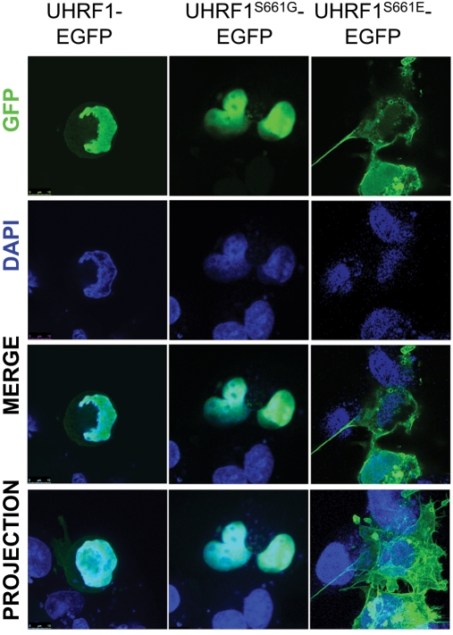
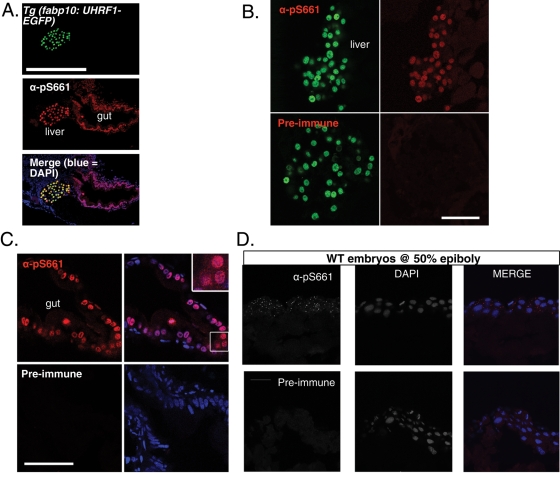
Ubiquitin-like, containing PHD and RING finger domains 1 (uhrf1) is regulated at the transcriptional level during the cell cycle and in developing zebrafish embryos. We identify phosphorylation as a novel means of regulating UHRF1 and demonstrate that Uhrf1 phosphorylation is required for gastrulation in zebrafish. Human UHRF1 contains a conserved cyclin-dependent kinase 2 (CDK2) phosphorylation site at Ser-661 that is phosphorylated in vitro by CDK2 partnered with cyclin A2 (CCNA2), but not cyclin E. An antibody specific for phospho-Ser-661 recognizes UHRF1 in both mammalian cancer cells and in nontransformed zebrafish cells, but not in zebrafish bearing a mutation in ccna2. Depleting Uhrf1 from zebrafish embryos by morpholino injection causes arrest before gastrulation and early embryonic death. This phenotype is rescued by wild-type UHRF1, but not by UHRF1 in which the phospho-acceptor site is mutated, demonstrating that UHRF1 phosphorylation is essential for embryogenesis. UHRF1 was detected in the nucleus and cytoplasm, whereas nonphosphorylatable UHRF1 is unable to localize to the cytoplasm, suggesting the importance of localization in UHRF1 function. Together, these data point to an essential role for UHRF1 phosphorylation by CDK/CCNA2 during early vertebrate development.
Figures
Similar articles
-
UHRF1 regulation of Dnmt1 is required for pre-gastrula zebrafish development.Dev Biol. 2016 Apr 1;412(1):99-113. doi: 10.1016/j.ydbio.2016.01.036. Epub 2016 Feb 3. Dev Biol. 2016. PMID: 26851214 Free PMC article.
-
Uhrf1 and Dnmt1 are required for development and maintenance of the zebrafish lens.Dev Biol. 2011 Feb 1;350(1):50-63. doi: 10.1016/j.ydbio.2010.11.009. Epub 2010 Nov 30. Dev Biol. 2011. PMID: 21126517 Free PMC article.
-
DNA hypomethylation induces a DNA replication-associated cell cycle arrest to block hepatic outgrowth in uhrf1 mutant zebrafish embryos.Development. 2015 Feb 1;142(3):510-21. doi: 10.1242/dev.115980. Epub 2015 Jan 6. Development. 2015. PMID: 25564650 Free PMC article.
-
Ca(2+)/calmodulin-dependent protein kinase phosphatase (CaMKP) is indispensable for normal embryogenesis in zebrafish, Danio rerio.Arch Biochem Biophys. 2009 Aug 1;488(1):48-59. doi: 10.1016/j.abb.2009.06.003. Epub 2009 Jun 13. Arch Biochem Biophys. 2009. PMID: 19527677
-
Hmx1 regulates urfh1 expression in the craniofacial region in zebrafish.PLoS One. 2021 Jan 19;16(1):e0245239. doi: 10.1371/journal.pone.0245239. eCollection 2021. PLoS One. 2021. PMID: 33465110 Free PMC article.
Cited by
-
Methylation of UHRF1 by SET7 is essential for DNA double-strand break repair.Nucleic Acids Res. 2019 Jan 10;47(1):184-196. doi: 10.1093/nar/gky975. Nucleic Acids Res. 2019. PMID: 30357346 Free PMC article.
-
Developmental Functions of the Dynamic DNA Methylome and Hydroxymethylome in the Mouse and Zebrafish: Similarities and Differences.Front Cell Dev Biol. 2018 Mar 20;6:27. doi: 10.3389/fcell.2018.00027. eCollection 2018. Front Cell Dev Biol. 2018. PMID: 29616219 Free PMC article. Review.
-
Targeted disruption of fibrinogen like protein-1 accelerates hepatocellular carcinoma development.Biochem Biophys Res Commun. 2015 Sep 18;465(2):167-73. doi: 10.1016/j.bbrc.2015.07.078. Epub 2015 Jul 28. Biochem Biophys Res Commun. 2015. PMID: 26225745 Free PMC article.
-
Structural insight into coordinated recognition of trimethylated histone H3 lysine 9 (H3K9me3) by the plant homeodomain (PHD) and tandem tudor domain (TTD) of UHRF1 (ubiquitin-like, containing PHD and RING finger domains, 1) protein.J Biol Chem. 2013 Jan 11;288(2):1329-39. doi: 10.1074/jbc.M112.415398. Epub 2012 Nov 16. J Biol Chem. 2013. PMID: 23161542 Free PMC article.
-
TGF-β signaling controls Foxp3 methylation and T reg cell differentiation by modulating Uhrf1 activity.J Exp Med. 2019 Dec 2;216(12):2819-2837. doi: 10.1084/jem.20190550. Epub 2019 Sep 12. J Exp Med. 2019. PMID: 31515281 Free PMC article.
References
-
- Abbady AQ, Bronner C, Bathami K, Muller CD, Jeanblanc M, Mathieu E, Klein JP, Candolfi E, Mousli M. TCR pathway involves ICBP90 gene down-regulation via E2F binding sites. Biochem Pharmacol. 2005;70:570–579. - PubMed
-
- Abbady AQ, Bronner C, Trotzier MA, Hopfner R, Bathami K, Muller CD, Jeanblanc M, Mousli M. ICBP90 expression is downregulated in apoptosis-induced Jurkat cells. Ann NY Acad Sci. 2003;1010:300–303. - PubMed
-
- Arima Y, Hirota T, Bronner C, Mousli M, Fujiwara T, Niwa S, Ishikawa H, Saya H. Down-regulation of nuclear protein ICBP90 by p53/p21Cip1/WAF1-dependent DNA-damage checkpoint signals contributes to cell cycle arrest at G1/S transition. Genes Cells. 2004;9:131–142. - PubMed
Publication types
MeSH terms
Substances
Grants and funding
LinkOut - more resources
Full Text Sources
Molecular Biology Databases
