. 2011 Nov;7(11):e1002350.
doi: 10.1371/journal.ppat.1002350. Epub 2011 Nov 3.

Fatal prion disease in a mouse model of genetic E200K Creutzfeldt-Jakob disease

Affiliations

Fatal prion disease in a mouse model of genetic E200K Creutzfeldt-Jakob disease

Yael Friedman-Levi et al. PLoS Pathog. 2011 Nov.

Erratum in

Retraction in

Abstract

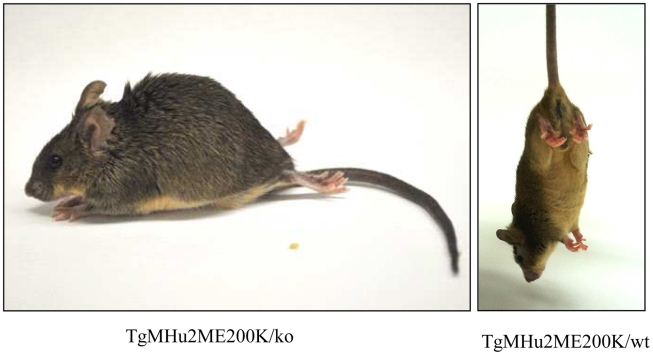
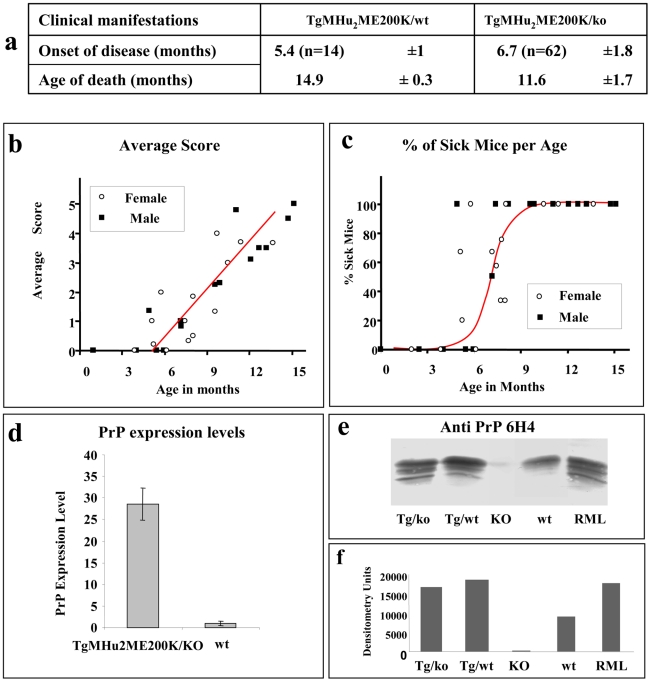
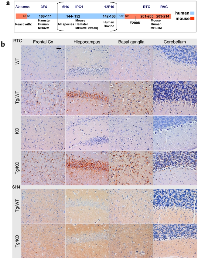
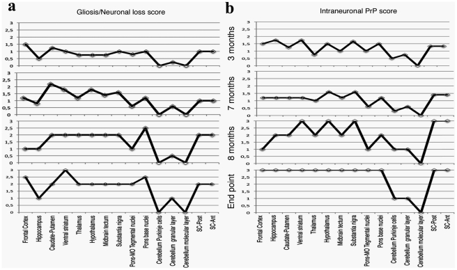
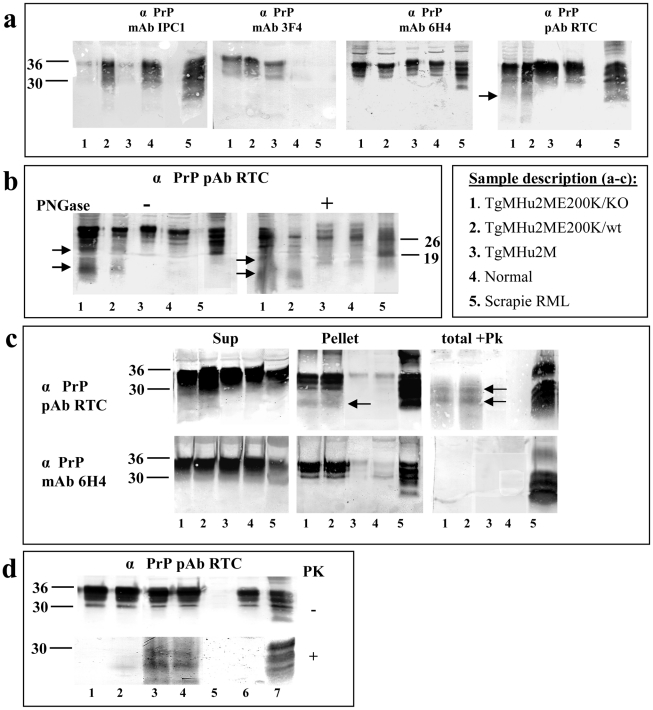
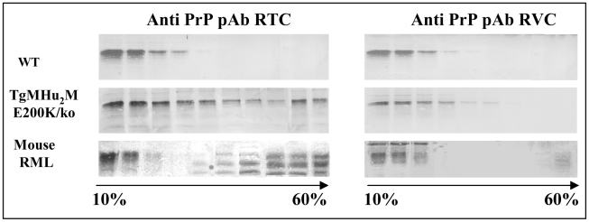
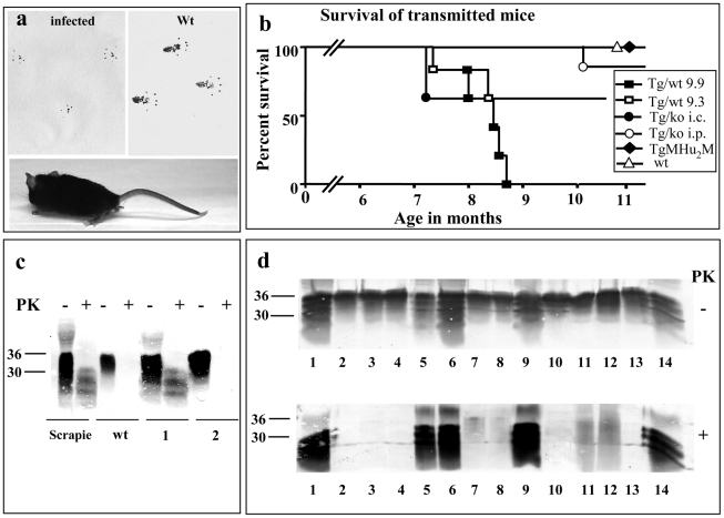
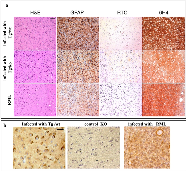
Genetic prion diseases are late onset fatal neurodegenerative disorders linked to pathogenic mutations in the prion protein-encoding gene, PRNP. The most prevalent of these is the substitution of Glutamate for Lysine at codon 200 (E200K), causing genetic Creutzfeldt-Jakob disease (gCJD) in several clusters, including Jews of Libyan origin. Investigating the pathogenesis of genetic CJD, as well as developing prophylactic treatments for young asymptomatic carriers of this and other PrP mutations, may well depend upon the availability of appropriate animal models in which long term treatments can be evaluated for efficacy and toxicity. Here we present the first effective mouse model for E200KCJD, which expresses chimeric mouse/human (TgMHu2M) E199KPrP on both a null and a wt PrP background, as is the case for heterozygous patients and carriers. Mice from both lines suffered from distinct neurological symptoms as early as 5-6 month of age and deteriorated to death several months thereafter. Histopathological examination of the brain and spinal cord revealed early gliosis and age-related intraneuronal deposition of disease-associated PrP similarly to human E200K gCJD. Concomitantly we detected aggregated, proteinase K resistant, truncated and oxidized PrP forms on immunoblots. Inoculation of brain extracts from TgMHu2ME199K mice readily induced, the first time for any mutant prion transgenic model, a distinct fatal prion disease in wt mice. We believe that these mice may serve as an ideal platform for the investigation of the pathogenesis of genetic prion disease and thus for the monitoring of anti-prion treatments.


Conflict of interest statement

The authors have declared that no competing interests exist.

Figures

Figure 1
Figure 1. Clinical Characterization of spontaneous disease in TgMHu2ME199K mice.
Typical pictures of sick TgMHu2ME199K mice showing hind limbs plegia, kyphosis and leg clasping.
Figure 2
Figure 2. Disease progression in spontaneous disease.
a: Average disease onset and death of mice in kinetic studies. b: Aggravation of clinical score of disease as related to the mice's age and gender. Groups of TgMHu2ME199K mice (male and female) were scored for clinical signs from birth to death. Average disease score for each group was plotted against the age elapsed since the mice birth. Closed circles: males, open circles: females. c: Percentage of sick mice in each age group. Groups of mice (as in fig b) in which the average score was at least 1 were plotted against the age of the mice. Closed circles: males, open circles: females d: Relative PrP mRNA levels, as determined by quantitative RT-PCR for wt and TgMHu2ME199K/ko mice. Each bar represents the average of PrP mRNA normalized against controls genes levels (see methods) in 4 male mice. Statistical bars represent standard error e: Brain homogenates from TgMHu2ME199K/ko, TgMHu2ME199K/wt, PrP ablated, wt C57BL/6 and RML-infected mice were immunoblotted with α PrP 6H4 mAb f: Relative intensities of the bands as measured by NIH Image J analysis software.
Figure 3
Figure 3. PrP immunoreactivity in the brains of TgMHu2ME199K mice.
a: Epitope mapping of α PrP antibodies used in these experiments. b: Four µm thick sections of formalin fixed, paraffin embedded brains from 8 months old TgMHu2ME199K on wt and ablated background, as compared to wt and PrP ablated mice were tested for disease related PrP immunoreactivity in diverse brain regions with both α PrP pAb RTC and α PrP mAb 6H4. The figure represents at least 3 samples from each group, and depicts intracellular PrP staining with RTC for the sick Tg mice. Scale bar in the upper left panel indicates 20 µm.
Figure 4
Figure 4. Summary of Pathology of TgMHu2ME199K mice: time course.
This figure summarizes the pathological findings in TgMHu2ME199K/ko mice of different ages (at least 3 mice for each age group, see Figure S1) a: the score for gliosis/neuronal loss in different brain areas of the TgMHu2ME199K mice as a function of aging. b: intraneuronal PrP immunoreactivity score in different brain areas as a function of aging.
Figure 5
Figure 5. Biochemical Characterization of PrP in TgMHu2ME199K mice.
a: To establish the PrP specificity of different samples, brain homogenates from 8 months old mice from 1: TgMHu2ME199K/ko, 2: TgMHu2ME199K/wt, 3: TgMHu2M, 4: wt C57BL/6, and 5: RML infected mice, were immunoblotted with several α PrP antibodies (See figure 3a for α PrP epitope mapping). Arrows demonstrate truncated PrP forms only present in brains of TgMHu2ME199K mice. b: Samples as in panel a were treated in the presence or absence of PNGase and immunoblotted with designated α PrP antibodies. As above truncated PrP forms are demonstrated only in the TgMHu2ME199K mice demonstrated with arrows. c: Samples as in panel a were extracted with sarkosyl and then centrifuged at 100000 g for 1 h, separated into pellets and supernatants. Otherwise, similar samples (denominated as total) were digested with 30 ug/ml PK for 30 min at 37°C. All samples were then immunoblotted with the designated anti PrP antibodies. Arrows demonstrate PK resistant bands in the TgMHu2ME199K samples. d: Samples from TgMHu2ME199K/ko mice at different ages and controls were digested in the presence or absence of 30 ug/ml PK for 30 min at 37°C and immunoblotted with α PrP pAb RTC. #1: 1 month old; #2: 3 months old; #3: 7 months old; #4: another 7 months old sample, #5 PrP ablated mouse; #6 wt mouse, #7 RML infected mouse.
Figure 6
Figure 6. Oxidation of TgMHu2ME199K PrP in Tg mice.
PrPSc in mice, hamsters and human samples cannot be detected by αPrP RVC, demonstrating it is oxidized in it helix3 Met residues. To see if this is also the case for all or part of E200K PrP in the Tg mice, brain homogenates from (wt C57BL/6, TgMHu2ME199K/ko, and scrapie RML), were extracted with sarkosyl and subjected to ultracentrifugation in 10–60% sucrose gradients. Individual fractions of each of the gradients were immunoblotted respectively with α PrP antibodies RTC and RVC. The figure shows that aggregated forms of the mutant protein are not recognized by this antibody, as is the case for RML PrPSc.
Figure 7
Figure 7. Transmission of TgMHu2ME199K prions to wt mice.
a: Picture of a wt C57BL/6 mouse infected with brain homogenates from TgMHu2ME199K/wt mice showing abnormal hind limb posture. Upper panel shows foot prints of the mice as compared to wt mice, demonstrating abnormal pattern of walking. b: Survival curves for wt C57BL/6 mice infected i.p with a TgMHu2ME199K/wt sick mouse brain (group 9.9, close square) or a TgMHu2ME199K/wt asymptomatic mouse brain (group 9.3, open square), as well as i.c (close circles) or i.p (open circles) infected with a TgMHu2ME199K/ko sick mouse brain, or i.p with the brain homogenate of TgMHu2M mice (close diamond) and i.c with wt brain homogenate (open triangle). c: Brain homogenates of RML infected and wt mice as well as mice (1&2) infected with TgMHu2ME199K/wt brain homogenate (group 9.3) immunoblotted in the presence and absence of PK digestion with α PrP pAb RTC. d: Brain homogenates digested in the presence or absence of PK and immunoblotted with α PrP pAb RTC of the brain samples described in table 3. Upper panel: −PK, lower Panel: +PK overdeveloped. Compare sample 1 in figures c and d to appreciate the overdevelopment factor.
Figure 8
Figure 8. Transmission of TgMHu2ME199K prions to wt mice: pathology.
a: Frontal cortex sections of mice infected with RML or with TgMHu2ME199K prions on a wt or ablated background, analyzed for prion pathological properties, spongiosis, gliosis, and disease related PrP immunoreactivity with pAb RTC and mAb 6H4. Scale bar in upper left image indicates 50 µm. b: RTC immunoreactivity in the absence of formic acid for brains infected with TgMHu2ME199K/wt or RML, as well as for naïve PrP ablated mice. Picture shows different pattern of immunoreactivity for both infected samples. Scale bar in upper left image indicates 20 µm.

References

    1. Kovacs GG, Puopolo M, Ladogana A, Pocchiari M, Budka H, et al. Genetic prion disease: the EUROCJD experience. Hum Genet. 2005;118:166–174. - PubMed
    1. Hsiao K, Prusiner SB. Inherited human prion diseases. Neurology. 1990;40:1820–1827. - PubMed
    1. Hsiao K, Meiner Z, Kahana E, Cass C, Kahana I, et al. Mutation of the prion protein in Libyan Jews with Creutzfeldt-Jakob disease. N Engl J Med. 1991;324:1091–1097. - PubMed
    1. Korczyn AD, Chapman J, Goldfarb LG, Brown P, Gajdusek DC. A mutation in the prion protein gene in Creutzfeldt-Jakob disease in Jewish patients of Libyan, Greek, and Tunisian origin. Ann N Y Acad Sci. 1991;640:171–176. - PubMed
    1. Lee HS, Sambuughin N, Cervenakova L, Chapman J, Pocchiari M, et al. Ancestral origins and worldwide distribution of the PRNP 200K mutation causing familial Creutzfeldt-Jakob disease. Am J Hum Genet. 1999;64:1063–1070. - PMC - PubMed

Publication types