. 2011 Nov;7(11):e1002351.
doi: 10.1371/journal.ppat.1002351. Epub 2011 Nov 3.

Anti-filarial activity of antibiotic therapy is due to extensive apoptosis after Wolbachia depletion from filarial nematodes

Affiliations

Anti-filarial activity of antibiotic therapy is due to extensive apoptosis after Wolbachia depletion from filarial nematodes

Frederic Landmann et al. PLoS Pathog. 2011 Nov.

Abstract

Filarial nematodes maintain a mutualistic relationship with the endosymbiont Wolbachia. Depletion of Wolbachia produces profound defects in nematode development, fertility and viability and thus has great promise as a novel approach for treating filarial diseases. However, little is known concerning the basis for this mutualistic relationship. Here we demonstrate using whole mount confocal microscopy that an immediate response to Wolbachia depletion is extensive apoptosis in the adult germline, and in the somatic cells of the embryos, microfilariae and fourth-stage larvae (L4). Surprisingly, apoptosis occurs in the majority of embryonic cells that had not been infected prior to antibiotic treatment. In addition, no apoptosis occurs in the hypodermal chords, which are populated with large numbers of Wolbachia, although disruption of the hypodermal cytoskeleton occurs following their depletion. Thus, the induction of apoptosis upon Wolbachia depletion is non-cell autonomous and suggests the involvement of factors originating from Wolbachia in the hypodermal chords. The pattern of apoptosis correlates closely with the nematode tissues and processes initially perturbed following depletion of Wolbachia, embryogenesis and long-term sterilization, which are sustained for several months until the premature death of the adult worms. Our observations provide a cellular mechanism to account for the sustained reductions in microfilarial loads and interruption of transmission that occurs prior to macrofilaricidal activity following antibiotic therapy of filarial nematodes.


Conflict of interest statement

The authors have declared that no competing interests exist.

Figures

Figure 1
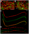
Figure 1. In vivo tetracycline treatment dramatically reduces the Wolbachia population in adult B. malayi females.
Female B. malayi lateral chords from un-treated (A) or tetracycline-treated (B) jirds. Total DNA is revealed with propidium iodide (red), host nuclei are counterstained with an anti acetylated histone H4 (green), therefore the red foci reveal only Wolbachia. The chords are flanked by muscle quadrants stained with phalloidin (green). Scale bar  = 100 µm.
Figure 2
Figure 2. Pyknotic nuclei and morphology defects observed in the germline and intrauterine microfilariae.
Germline cells-containing ovaries (A, B) and microfilariae obtained from uteri (C, D) were stained for DNA (propidium iodide, red) and actin (phalloidin, green). Worms dissected from un-treated animals (A, C), and from animals treated with tetracycline (B, D). In (A), propidium iodide reveals germline nuclei surrounded by Wolbachia (appearing as small red foci), while Wolbachia are absent in (B). In (B), arrowheads point towards some pyknotic nuclei, with a condensed chromatin appearing brighter with propidium iodide stain, while stars indicate germ cell nuclei with a normal morphology. Note that depending on the area observed in the ovary, wild type nuclei can slightly vary in volume, as shown in images (A) and (B). Scale bar  = 20 µm.
Figure 3
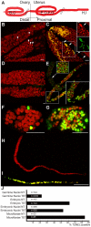
Figure 3. In vivo tetracycline treatment leads to apoptosis in adult worm reproductive tissues.
(A) Schematic drawing of one female reproductive tract, representing the approximate localization of the different sections observed in the TUNEL assay. TUNEL experiments showing the DNA (PI in red) and incorporated fluorescein-dUTP (green), in samples from non-treated (B, D, F, H) or tetracycline-treated (C, E, G, I) B. malayi females. Mitotic proliferation zone in the distal ovary (B, C). Arrowheads indicate mitotic nuclei, arrows point to somatic gonad nuclei. (D) Uterus filled with elongating embryos. (E) Fertilization area in the distal uterus in the top left corner, and proximal uterus filled (in diagonal) with developing embryos. (F, G) Single developing embryos. (H, I) Intrauterine microfilariae extracted from proximal uteri. (J) TUNEL quantification. For each un-treated (NT) or tetracycline-treated (TET) sample, TUNEL-positive nuclei or embryos were counted and expressed as a percentage of total nuclei or embryos (based on DNA staining with PI). For the embryonic count (“embryos NT”, “embryos TET”), embryos with a number of TUNEL positive nuclei equal or less than 2 positive nuclei were considered as negative embryos. All the intrauterine microfilariae found TUNEL-positive in the TET sample had every nuclei TUNEL-positive (I). Scale bar  = 15 µm.
Figure 4
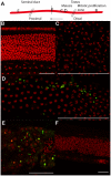
Figure 4. Tetracycline treatment induces detectable apoptosis during spermatogenesis.
TUNEL experiments showing the DNA (PI in red) and incorporated fluorescein-dUTP (green), in samples from un-treated (B, C) or tetracycline-treated (D to F) B. malayi males. (A) Schematic drawing of the male reproductive tract, representing the approximate localization of the different sections observed in the TUNEL assay. (B) Mitotic proliferation zone of spermatogoniae. (C, D) Synaptonemal complexes in meiosis I. (E) Proximal testes. (F) Seminal duct filled with mature spermatocytes. Scale bar  = 100 µm.
Figure 5
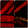
Figure 5. Cytoskeleton defects are revealed in somatic tissues, without apoptotic phenotypes.
Female B. malayi lateral chords from un-treated (A, C) or tetracycline-treated (B, D) jirds. No pyknotic nuclei were detected, and the TUNEL levels were similar in both samples (A, B). (C, D) Apical microtubule network. Scale bar  = 100 µm.
Figure 6
Figure 6. Doxycycline treatment leads to apoptosis in vitro.
(A, C) control worms and treated worms (B, D) were TUNEL assayed (green) and stained for DNA (PI in red). (A, B) Germ cells in mitotic proliferation in the ovaries. (C) Proximal uteri filled with developing embryos. (D) Apoptotic oocytes and early embryos (arrows) in a distal uterus, surrounded by sperm cells (arrowheads). Scale bar  = 15 µm.
Figure 7
Figure 7. Increased expression of ced-3 gene and activation of CED-3 protein in tetracycline treated B. malayi.
A) Ced-3 gene expression level normalized by expression level of gst in BM (B. malayi) and BM-TET (tetracycline treated B. malayi) adult females. B) Western blot detection of CED-3 in microfilaria and 14 day old L4 larvae. Lanes 1-2, microfilariae from untreated control (1) and tetracycline treated (2). Lanes 3-4 lanes, L4 larvae, untreated control (3) and tetracycline treated (4). Activated (cleaved) CED-3 is more abundant in microfilariae and L4 larvae from treated jirds compared to untreated controls. Lane 5, molecular weight markers.
Figure 8
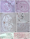
Figure 8. Apoptosis and apoptotic bodies are detected in O. volvulus tissues from human nodules of doxycycline treated patients.
A, B. Cross-sections of adult female worm showing absence of apoptosis and intact embryonic inter-uterine stages (oocytes, pretzel stages, coiled embryonic microfilariae). C-G. Cross-sections of adult female worms depleted of Wolbachia showing extensive apoptosis of germline and early embryonic cells and uterine epithelial cells. Stars label inter-uterine content, black arrowheads label apoptotic germline and early embryonic cells as well as human cells surrounding the worm, white arrowheads point to somatic cells, such as epithelial cells surrounded uteri. Scale bar  = 20 µm.
Figure 9
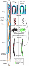
Figure 9. Wolbachia-dependent non cell-autonomous apoptosis.
Schematic drawing of a B. malayi female focusing on the reproductive apparatus, showing levels of apoptosis before and after Wolbachia removal. Apoptosis remains a rare event during germline maturation, and is developmentally programmed during embryogenesis. After Wolbachia depletion, cumulative apoptosis is observed in germ cell, embryo and microfilaria. The absence of Wolbachia (from the hypodermal chords and from the few embryonic cells derived from the C blastomere) leads to a massive non cell-autonomous, “bystander” apoptosis, in embryonic cells normally devoid of Wolbachia (green nuclei).

References

    1. Kozek WJ. Transovarially-transmitted intracellular microorganisms in adult and larval stages of Brugia malayi. J Parasitol. 1977;63:992–1000. - PubMed
    1. Kozek WJ, Marroquin HF. Intracytoplasmic bacteria in Onchocerca volvulus. Am J Trop Med Hyg. 1977;26:663–678. - PubMed
    1. Taylor MJ, Hoerauf A. Wolbachia bacteria of filarial nematodes. Parasitol Today. 1999;15:437–442. - PubMed
    1. Taylor MJ, Bilo K, Cross HF, Archer JP, Underwood AP. 16S rDNA phylogeny and ultrastructural characterization of Wolbachia intracellular bacteria of the filarial nematodes Brugia malayi, B. pahangi, and Wuchereria bancrofti. Exp Parasitol. 1999;91:356–361. - PubMed
    1. Taylor MJ, Hoerauf A, Bockarie M. Lymphatic filariasis and onchocerciasis. Lancet. 2010;376:1175–1185. - PubMed

Publication types

MeSH terms