. 2011 Nov;7(11):e1002358.
doi: 10.1371/journal.pgen.1002358. Epub 2011 Nov 3.

A novel protein LZTFL1 regulates ciliary trafficking of the BBSome and Smoothened

Affiliations

A novel protein LZTFL1 regulates ciliary trafficking of the BBSome and Smoothened

Seongjin Seo et al. PLoS Genet. 2011 Nov.

Abstract

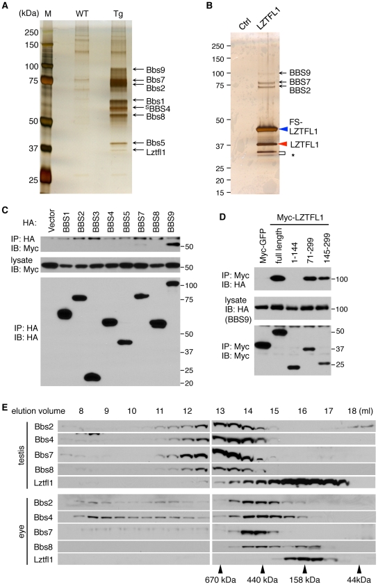
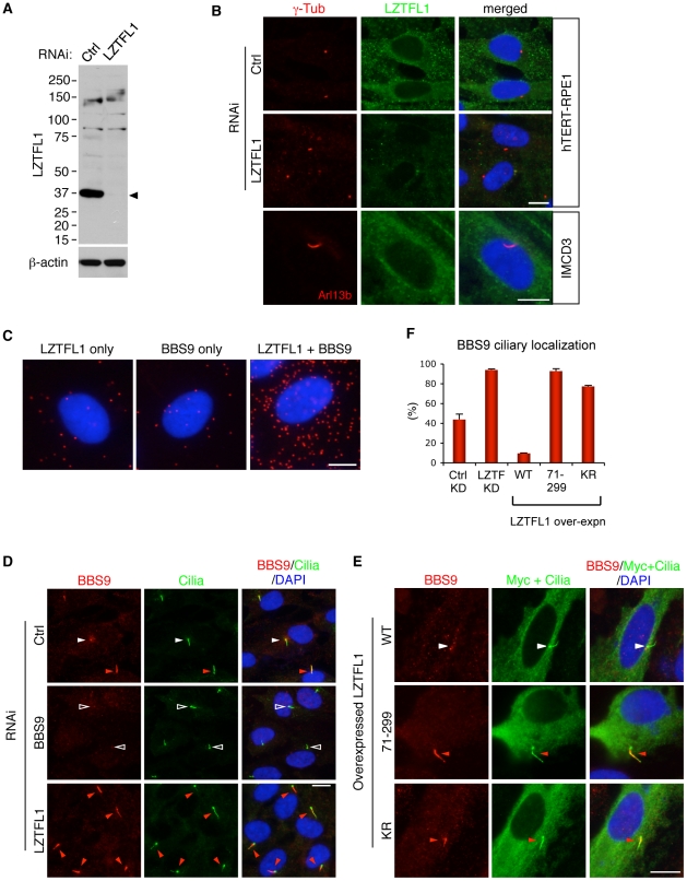
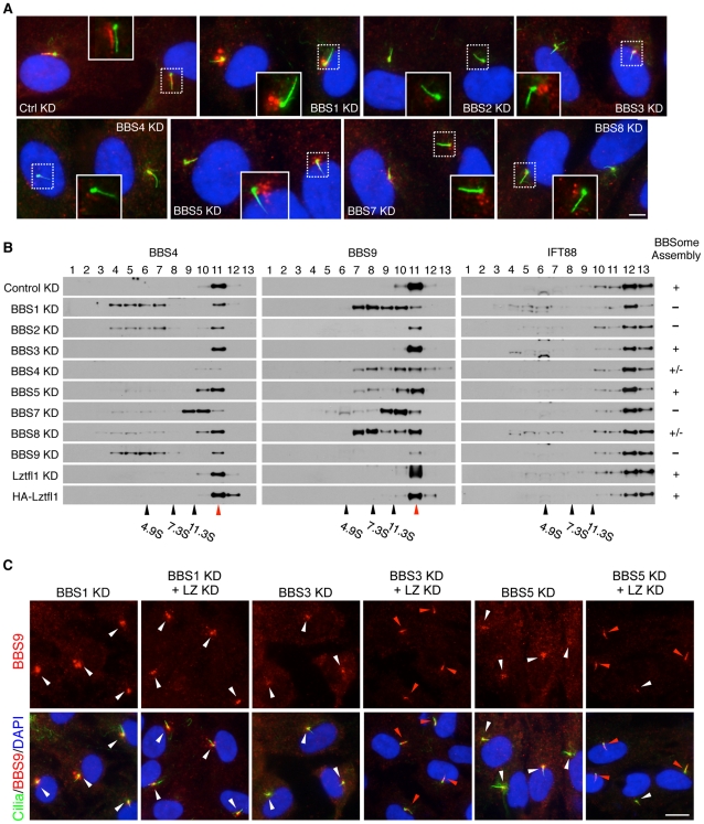
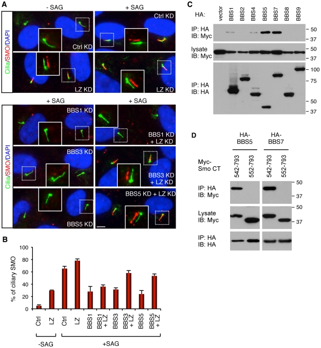
Many signaling proteins including G protein-coupled receptors localize to primary cilia, regulating cellular processes including differentiation, proliferation, organogenesis, and tumorigenesis. Bardet-Biedl Syndrome (BBS) proteins are involved in maintaining ciliary function by mediating protein trafficking to the cilia. However, the mechanisms governing ciliary trafficking by BBS proteins are not well understood. Here, we show that a novel protein, Leucine-zipper transcription factor-like 1 (LZTFL1), interacts with a BBS protein complex known as the BBSome and regulates ciliary trafficking of this complex. We also show that all BBSome subunits and BBS3 (also known as ARL6) are required for BBSome ciliary entry and that reduction of LZTFL1 restores BBSome trafficking to cilia in BBS3 and BBS5 depleted cells. Finally, we found that BBS proteins and LZTFL1 regulate ciliary trafficking of hedgehog signal transducer, Smoothened. Our findings suggest that LZTFL1 is an important regulator of BBSome ciliary trafficking and hedgehog signaling.


Conflict of interest statement

The authors have declared that no competing interests exist.

Figures

Figure 1
Figure 1. Identification of LZTFL1 as a BBSome interacting protein.
(A) Co-purification of Lztfl1 as a BBSome-interacting protein from LAP-BBS4 transgenic mouse testis. Lysates from WT and Tg testes were subjected to TAP and purified proteins were separated by SDS-PAGE and silver-stained. Protein size markers (M) were loaded in the left lane. (B) FS-LZTFL1 (blue arrowhead) and its associated proteins were isolated by TAP from HEK293T cells and visualized by silver staining. Parental cells were used as a control. Red arrowhead indicates endogenous LZTFL1, and asterisk, two smaller forms of LZTFL1. (C) LZTFL1 interacts with BBS9 within the BBSome. Each subunit of the BBSome (HA-tagged) was co-transfected with Myc-tagged LZTFL1 into HEK293T cells and lysates were analyzed by co-immunoprecipitation (IP) using anti-HA antibodies. Bottom panel shows immunoprecipitated BBS proteins and middle, Myc-LZTFL1 in the lysates. The top panel shows Myc-LZTFL1 precipitated by anti-HA antibodies. (D) BBS9 binds to the C-terminal half of LZTFL1. Full-length and several deletion mutant variants of Myc-LZTFL1 were co-transfected with HA-BBS9 in HEK293T cells. Myc-GFP was used as a negative control and anti-Myc antibody was used for IP. Numbers indicate expressed portions of LZTFL1 in amino acid positions. (E) The vast majority of Lztfl1 is not associated with the BBSome. Lysates from WT mouse testis and eye were subjected to size exclusion chromatography using Superose-6 10/300 GL column. Eluted fractions were analyzed by SDS-PAGE followed by immunoblotting against indicated antibodies. Elution volume of protein standards is shown at the bottom.
Figure 2
Figure 2. LZTFL1 regulates ciliary trafficking of the BBSome.
(A) Verification of the specificity of anti-LZTFL1 antibody and siRNA-mediated knock-down of LZTFL1 expression in hTERT-RPE1 cells. Arrowhead depicts the LZTFL1 protein band. β-actin was used as a loading control. (B) LZTFL1 localizes to the cytoplasm but not cilia or basal body. Localization of LZTFL1 (green) was probed with anti-LZTFL1 antibody in hTERT-RPE1 (top and middle rows) and IMCD3 (bottom row) cells after 30 hrs of serum withdrawal. Antibodies against γ-tubulin and Arl13b (red) were used to mark the basal body and cilia, respectively. Nuclei were stained with 4',6-diamidino-2-phenylindole (DAPI, blue). (C) LZTFL1-bound BBSome localizes to the cytoplasm. Left and middle panels are negative controls (background), where only either LZTFL1 or BBS9 antibodies was used. In the right panel, both LZTFL1 and BBS9 antibodies were used. Red dots (“blobs”) represent the protein complexes containing both LZTFL1 and BBS9 detected by in situ Proximity-mediated Ligation Assay (PLA) in hTERT-RPE1 cells. (D) BBS9 ciliary localization increases in LZTFL1 depleted cells. In ciliated RPE1 cells, BBS9 (red) shows two distinct localization patterns: ciliary (red arrowhead) and peri-centriolar (white arrowhead). Open arrowhead depicts the lack of BBS9 in BBS9 siRNA transfected cells. Cilia (green) were marked by antibodies against acetylated tubulin and γ-tubulin. (E) Over-expression of LZTFL1 inhibits ciliary entry of BBS9. Myc-tagged LZTFL1 variants were transfected into RPE1 cells and BBS9 localization (red) was probed. Transfected cells were determined by using anti-Myc antibody (green). Scale bars, 10 µm. (F) Quantitation of BBS9 ciliary localization. The number of ciliated cells with ciliary BBS9 staining was counted. Results of knock-down (KD) experiments are average of four independent experiments with at least 100 cells counted in each experiment. Over-expression results are the average of two independent experiments with at least 40 cells counted in each experiment. Data are shown as means ± SEM.
Figure 3
Figure 3. Reduction of LZTFL1 activity restores BBSome ciliary trafficking in BBS3 and BBS5 depleted cells.
(A) All BBSome subunits are required for BBSome ciliary entry. RPE1 cells were transfected with siRNAs against each BBSome subunit and BBS3. Cilia and basal body are labeled with acetylated tubulin and γ-tubulin antibodies (green) and BBS9 is in red. The inlets are shifted overlay images of the boxed area. Scale bar, 5 µm. (B) BBSome status in the absence of individual BBSome components. Proteins from hTERT-RPE1 cells transfected with siRNAs against each BBS gene were separated by 10–40% sucrose gradient centrifugation and fractions were analyzed by immunoblotting using antibodies against BBS4, BBS9 and IFT88. Impact of LZTFL1 depletion and over-expression on BBSome assembly was also examined. Migration of molecular weight standards is shown at the bottom and red arrowhead indicates intact BBSome (14S). The BBSome assembly status was ranked by the amount of remaining intact BBSome (except for the depleted subunit) and summarized in the right. +: Normal BBSome assembly (more than 70% remaining), +/−: moderate decrease (30–70% remaining) severe decrease (less than 30% remaining). (C) Restoration of BBSome ciliary trafficking in BBS3 and BBS5 depleted cells by LZTFL1 depletion. RPE1 cells were transfected as indicated, and BBS9 (red) localization was examined. Scale bar, 10 µm.
Figure 4
Figure 4. BBSome and LZTFL1 regulate SMO ciliary trafficking.
(A) BBSome is required for SMO ciliary localization, while LZTFL1 inhibits it. hTERT-RPE1 cells were depleted with BBSome subunits and LZTFL1 (LZ) by RNAi. After 24 hrs of serum withdrawal, cells were incubated with or without SMO agonist (SAG) for 4 hrs. Cells were labeled with antibodies to acetylated tubulin and γ-tubulin (green) and SMO (red). Scale bar, 5 µm. (B) Quantitation of SMO ciliary localization. Graphs are average percentages of SMO positive cilia from four independent experiments with minimum 90 cells counted in each experiment. Data are shown as means ± SEM. (C) Physical interaction between BBSome and Smo C-terminal cytoplasmic tail domain. Indicated BBSome subunits (HA-tagged) were co-transfected with Myc-tagged Smo cytoplasmic tail domain (aa 542–793) and lysates were subjected to co-IP using anti-HA antibodies. (D) Deletion of 10 amino acids, which contains the WR motif, from the N-terminus of Smo cytoplasmic tail domain abolishes the interaction between Smo and BBS proteins.

References

    1. Singla V, Reiter JF. The primary cilium as the cell's antenna: signaling at a sensory organelle. Science. 2006;313:629–633. - PubMed
    1. Goetz SC, Anderson KV. The primary cilium: a signalling centre during vertebrate development. Nat Rev Genet. 2010;11:331–344. - PMC - PubMed
    1. Quinlan RJ, Tobin JL, Beales PL. Modeling ciliopathies: Primary cilia in development and disease. Curr Top Dev Biol. 2008;84:249–310. - PubMed
    1. Badano JL, Mitsuma N, Beales PL, Katsanis N. The ciliopathies: an emerging class of human genetic disorders. Annu Rev Genomics Hum Genet. 2006;7:125–148. - PubMed
    1. Zaghloul NA, Katsanis N. Mechanistic insights into Bardet-Biedl syndrome, a model ciliopathy. J Clin Invest. 2009;119:428–437. - PMC - PubMed

Publication types

MeSH terms