. 2004:3:Doc02.
Epub 2004 Dec 28.

Cellular and molecular mechanisms in environmental and occupational inhalation toxicology

Affiliations

Cellular and molecular mechanisms in environmental and occupational inhalation toxicology

Herbert Riechelmann. GMS Curr Top Otorhinolaryngol Head Neck Surg. 2004.

Abstract

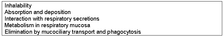
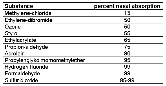
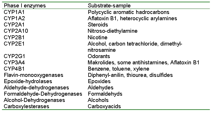
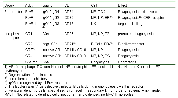
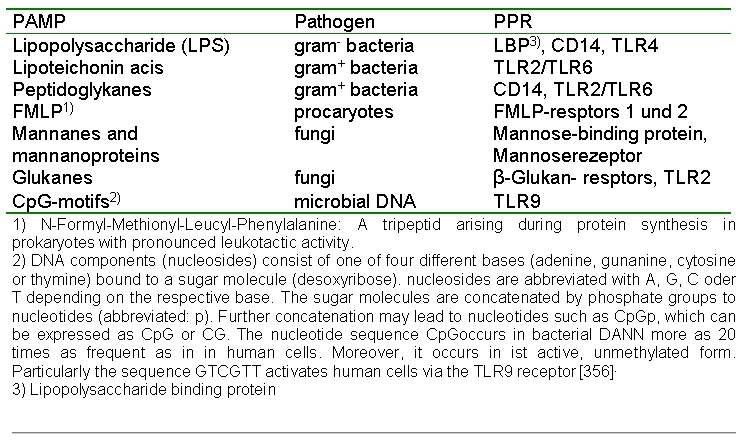
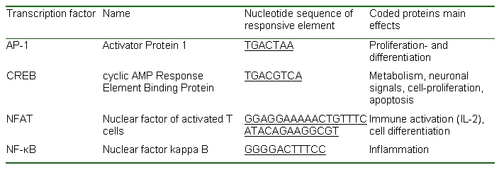
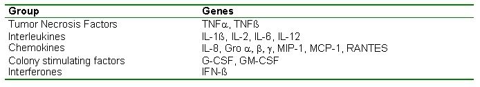
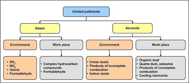
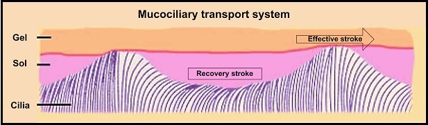
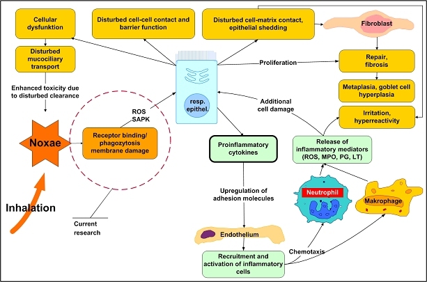
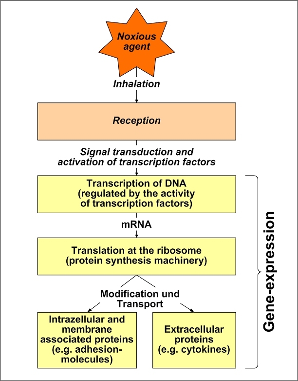
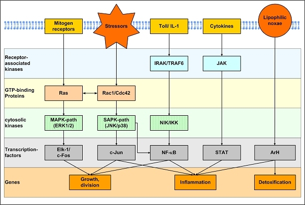
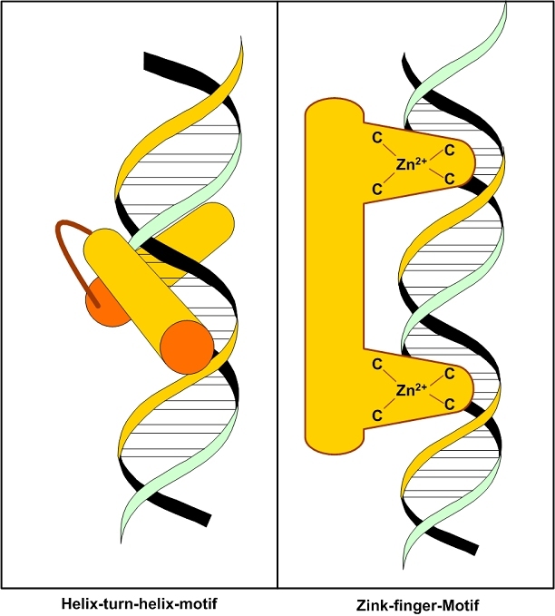
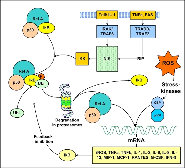
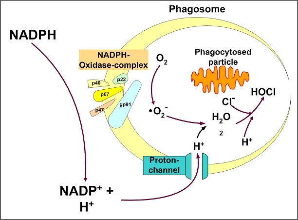
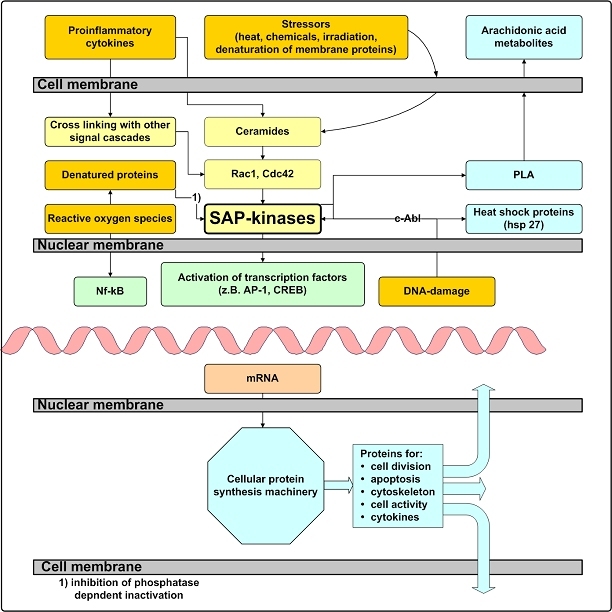
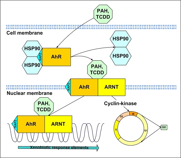
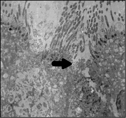
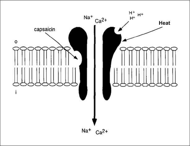
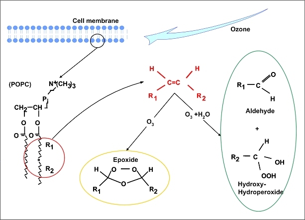
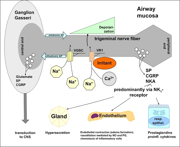
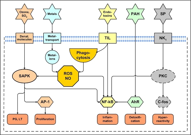
The central issue of this review are inflammatory changes that take place in the mucous membranes of the respiratory tract as a result of inhaled pollutants. Of particular relevance are dusts, SO(2), ozone, aldehydes und volatile organic compounds. Bioorganic pollutants, especially fragments of bacteria and fungi, occur predominantly in indoor dusts. They activate the toll-like/IL-1 receptor and lead to the activation of the transcription factor NF-κB for the release of numerous proinflammatory cytokines. Metals are predominant in ambient air dust particles. They induce the release of reactive oxygen species that cause damage to lipids, proteins and the DNA of the cell. As well as NF-κB, transcription factors that foster proliferation are activated via stress activated protein kinases. Organic compounds such as polycyclic aromatic hydrocarbons and nitroso-compounds of incomplete combustion processes activate additional via the cytosolic arylhydrocarbon receptor for detoxification enzymes. Sulphur dioxide leads to acid stress, and ozone to oxidative stress of the cell. This is accompanied by the release of proinflammatory cytokines via stress activated protein kinases. Aldehydes and volatile organic compounds activate the vanilloid receptor of trigeminal nerve fibres and induce a hyperreactivity of the mucous membrane via the release of nerve growth factors. The mechanisms described work synergistically and lead to a chronic inflammatory reaction of the mucous membranes of the upper respiratory tract that is regularly demonstrable in inhabitants of western industrial nations. It is unclear whether we are dealing here with a physiological inflammation or with an at least partially avoidable result of chronic pollutant exposure.

Keywords: cell membrane receptors; endotoxins; metals; nitrogen monoxide; polycyclic aromatic hydrocarbons; reactive oxygen species; respiratory mucous membrane; signal transduction; toxicology; transcription factors.


Figures

Table 1
Table 1. Toxicokinetic factors in respiratory toxicology
Table 2
Table 2. Absorption of gaseous pollutants in upper airways
Table 3
Table 3. Phase I enzymes detected in the upper airway mucosa
Table 4
Table 4. Characteristics of Fc- and complement-receptors
Table 5
Table 5. Pathogen-associated molecular patterns (PAMP) on particle-adsorbed bioorganic pollutants, related pathogens and pattern recognition receptors (PPR)
Table 6
Table 6. Some nucleotide sequences of responsive elements recognized by transcription factors [357-360]
Table 7
Table 7. Gene transcription regulated by NF-κB
Figure 1
Figure 1. Intrinsic and extrinsic factors in the development of disease
Figure 2
Figure 2. Relevant environmental and occupational inhalative pollutants
Figure 3
Figure 3. Particle deposition according to ISO 7708
The inhaled fraction and the percentage deposited in various airway compartments is depicted in percent dependant on mean mass aerodynamnic diameter.
Figure 4
Figure 4. Radiography of a whole body section of the rat after intravenous injection of radioactive 1,2-dibromo-ethane
High metabolic activity is found in the liver and in the mucosa of the upper and lower airways.
Figure 5
Figure 5. Metabolism of benzene
A phase reaction results in the formation of a highly toxic itermediate product benzene epoxide, which is conjugated to to glucurnic acid to form a less toxic product, which can be renaly excreted.
Figure 6
Figure 6. Mucociliary transport system of the upper airways
Cilia xxxxx in the watery periciliary fluid and grip into and propel the superficial mucus blanket during the effective stroke. The mucus blanket is thus moved to the nasopharynx, from where it is swallowed together with adherent pollutants.
Figure 7
Figure 7. In the cells of the upper airway mucosa, inhalative pollutanta induce the release of proinflammatory cytokines.
They cause endothelial cells in neighboring vessels to express adhesion molecules in their cell membrane. Infalmmatory cells from the blood stich to those adhesion molecules and are transferred to the interstitial tissues and airway epithelium. Here, these cells release further inflammatory mediators including ROS, myeloperoxidase (MPO), prostaglandins (PG) and leukotrienes (LT). These mediators induce cellular dysfunction, disturbes cell-cell contacts and shedding of cells from the basal membrane. Sensible nerve endings are uncovered promoting airway hyperreactivity. Tissue repair promoted by fibroblasts may lead to structural changes with exaggerated production of collagen fibers rsulting in basal membrane thickening (remodelling). If inflammation persists, regenerated cells do not differentiate into ciliated, but into squamous epithelium (metaplasia) and goblet cells (goblet cell hyperplasia). Current research focuses on the mechanisms which induce the release of proinflammatory cyokines from airway cells (red circle). Pollutant binding to cellular receptors and and the concomitant release of ROS are supposed key mechansisms. ROS and additional intermediates damage the cells membrane and activate intracellular signal transduction cascades (stress activated protein kinases - SAPK) finally resulting in the release of cytokines. Not depicted are specific immune responses and changes due to DNA damage.
Figure 8
Figure 8. Altered gene expression following contact with an inhalative pollutant
By various mechanisms, the cell detects the inhaled pollutant (perception). This generates an intracellular signal, which is transferred by different cellular transduction systems and converge to transcription factors. Transcription factors are then transferred to the cell nucleus, where they bind to specific DNA-regions modifying the transcription into pre-mRNA. Following posttranscriptional modification, the resulting mature RNA leaves the nucleus into the cytoplasm, where the translation into a protein at the reibosoes takes place. The generated proteins can be excretes from the cell, integrate into the cell membrane to modify cell reactivity or serve as intracellular proteins various cell functions.
Figure 9
Figure 9. The MAP-Kinase pathway, SAP-Kinasae, the activation of NF-κB following activation of the Toll-like/IL-1 Receptor, and activation of STAT via cytokine-receptors as paradigms of intracellular signal trandusction mechanisms (s. text).
Lipophilic noxae can pass the cell membrane and activate the cytosolic arylhydrocarbon-receptor (AhR) directly, which then serves as a transcription factor.
Figure 10
Figure 10. Strucutral motifs of the DNA-binding domain of regulatory transcription factors fit into grooves of the DNA molecule.
Depicted are the Helix-Turn-Helix-Motif and the Zink-Finger-Motif.
Figure 11
Figure 11. Activation of NF-κB
Ligand binding to different cell membrane receptors (Toll-like/IL-1, TNF) activates intracellular signal transduction molecules such as the receptor associated protein kinases Interleukin 1 Receptor-Associated Kinase (IRAK) and TNF receptor-associated factor (TRAF) and then the cytosolic proteinkinase NF-κB-inducing Kinase (NIK). This kinase phophorlyses IkB Kinase (IKK), which in turn activates IκB. Phosphorylsed IκB binds to Ubiquitin, which transfers the NF-κB/IκB-complex to cellular proteasomes for degradation. Following Abspaltung of IκB within the proteasome, NF-κB can be transferred into the nucleus and promote the transcription of genes with appropriate responsive elements. Reaktive oxygen species activate stress-activated-protein-kinases, which in turn activate auxiliary proteins such as CBP and p300. This enhances the NF-κB binding to its responsive elements.
Figure 12
Figure 12. During phagocytosis, intracellular reactive oxygen species rapidly increase, known as oxidative burst.
Following receptor binding od , e.g. LPS, and particle internalization, the protein p47phox is activated and integrates with the additional components p67phox and p40phox into the phagosome membrane, where it combines with flavocytochorme b to the active NADPH-oxidase complex. This enzyme complex catalyzes the generation of ROS and protons, which shift through proton-channels into the interior of the phagosome, where they destroy the internalized particle.
Figure 13
Figure 13. The stress activated protein-kinase path
SAP kinases are a group of cytosolic protein kinases activated in a cascade like fashion following cellular stress exposure. Their activation occurs basically via 4 mechanisms. Proimflammatory cytokines and growth factors activate SAP-kinases via interconnections with their main intracellular pathways mediated by small GTP binding proteins such as Rac1 or Cdc42. Extracellular stressors cause the generations of cermides in the within the cellular lipid membrane, which activate SAP-kinases as second messengers. DNA damage activates the tyrosinkinase c-Abl, which activates SAP-kinases via intermediary steps. Intracellular ROS denature phosphatases and thus interfere with the inactivation of SAP-kinases. In addition, ROS activate the transcriptionfactor NF-κB by mechanisms not yet understood. Activation of SAP kinases in turn activates several transcription factors, which then translocate into the nucleus. Here they activates genes coding for proteins involved in cell division, apoptosis, cytoskeleton, cell activity and inflammation. In addition, SAP-kinases activate phsopholipase A2 thus altering the release of prostaglandins and leukotrienes. Heat shock proteins belong to the chaperones, which repair denatured proteins (resoration of their three-dimensional structure). Heat skock proteins serve as an indicator for cellular stress.
Figure 14
Figure 14. The arylhydrocarbon receptor (AhR) belongs to the Ligand activated basic helix-loop-helix transcription factors.
In the cytosol, it occurs as an tetrameric complex with heat shock protein (HSP) 90 and X-associated protein 2 (syn: ARA9). Ligands such as dioxin (TCCD) or polycyclic aromatic hydrocarbons dissolve the 2 HSP molecules from the complex. The remaining complex binds to the Arylhydrocarbon Receptor Nucleus Translocator (ARNT), is translocated in to the nucleus and enhance the transcription of xenobiotic response element dependent genes. The gene expression of several proteins including various detoxification enzymes is increased. In addition, cyclin kinase 2 is activated and TGF-β effects are inhibited, resulting in increased cell division.
Figure 15
Figure 15. Vacuolization and interruption of tight junctions (arrow) in a guinea pig trachea following exposure to 7,5 mg/m3 SO2
Figure 16
Figure 16. Proton activation of vanilloid-receptors
Vanilloid such as Capsaicin, various environmental irritants and heat (> 43°C) may open the Vanilloid receptor for Na+ and Ca2+ ions, whereas H+-Ions are not able to activate the receptor. However, protons bind to the receptor and increase its responsiveness to protons and irritants (permissive activity).
Figure 17
Figure 17. Peroxidation of membrane lipids (here: 1-palmitoyl-2-oleyl-sn-glycerol-3-phosphocholin (POPC)) and generation of lipid ozonation products by inhaled ozone attacks the double-bound in the right fatty acid (red circle) and epoxides it to an ozonide (yellow circle) or hydrolyzes it to an aldehyde and a hydroxyhydroperoxide (right, green circle).
Figure 18
Figure 18. Mucosal irritation and neurogenic inflammation
An irritant binds to a vanilloid type I receptor (VR1) on a trigeminal, non-myelinated group C fiber within the airway mucosa (nociceptor). Local influx of sodium and calcium ions results in circumscribed membrane depolarization, which in turn opens voltage gated sodium channels (VGSC) and generates an action potential along the nerve fiber. This action potential runs orthodromically (in the correct direction) toward the Gasserian ganglion, where transmitter substances including glutamate, Substance P (SP) und Calcitonin Gene Related Peptide (CGRP) transfers the signal to central neurons responsible for the perception of irritation and pain. The action potential also runs antidromically toward the periphery, where tachykinins such as SP, CGRP and Neurokinin A (NKA) are released. They activate the Neurokinin 1 (NK1) receptor on glandular cells, respiratory epithelium and endothelium of neighbouring vessels. Thus they mediate hypersecretion, vasodilation and inflammatory cell infiltration.
Figure 19
Figure 19. Synopsis of frequent toxicity mechanisms of inhaled pollutants
Ozone and SO2 denature membrane components, which activate stress activated protein kinases (SAPK). This results in the release of prostaglandins (PG) and leukotrienes (LT) and in the activation of transcriptionfactors such as C-jun, which activates among others genes for cell proliferation. SAPK also activate NF-κB, a key transcription factor inducing inflammatory cell reactions. Metals are internalized via cellular metal transporter proteins and induce the release of reactive oxygen species (ROS) and nitrogen monoxide (NO), which in turn activate SAPK and NF-κB. Bioorganic pollutants bind to the Toll-like/IL-1 (TIL-) receptor and activate NF-κB via cytosolic signal transduction pathways. In addition, particles with adsorbed bioorganic pollutants are phagocytosed and induce the release of ROS and NO via the respiratory burst. Lipophilic pollutants such as polycyclic aromatic hydrocarbons (PAH) pass the cell membrane and bind to the cytosolic arylhydrocarbon receptor (ArH), which in addition to NF-κB activates genes for detoxification enzymes. Irritants release substance P from nociceptors, which bind to the neurokinin receptor 1 (NK1) and activate genes for nerve growth factors (neurotrophins), probably via activation of Proteinkinase C (PKC) and the transcription factor C-fos. This may result in airway hyperreactivity. In addition, NF-κB is activated via the NK1-receptor.

References

    1. von-Mutius E, Sherrill DL, Fritzsch C, Martinez FD, Lebowitz MD. Air pollution and upper respiratory symptoms in children from East Germany. Eur Respir J. 1995;8:723–728. - PubMed
    1. Calderon-Garciduenas L, Rodriguez-Alcaraz A, Villarreal-Calderon A, Lyght O, Janszen D, Morgan KT. Nasal epithelium as a sentinel for airborne environmental pollution. Toxicol Sci. 1998;46:352–364. - PubMed
    1. Keles N, Ilicali OC, Deger K. Impact of air pollution on prevalence of rhinitis in Istanbul. Arch Environ Health. 1999;54:48–51. - PubMed
    1. Keles N, Ilicali C. The impact of outdoor pollution on upper respiratory diseases. Rhinology. 1998;36:24–27. - PubMed
    1. Abbey DE, Hwang BL, Burchette RJ, Vancuren T, Mills PK. Estimated long-term ambient concentrations of PM10 and development of respiratory symptoms in a nonsmoking population. Arch Environ Health. 1995;50:139–152. - PubMed

LinkOut - more resources