. 2005:4:Doc01.
Epub 2005 Sep 28.

Therapy of hearing disorders - conservative procedures

Affiliations

Therapy of hearing disorders - conservative procedures

Stefan Plontke. GMS Curr Top Otorhinolaryngol Head Neck Surg. 2005.

Abstract

A wide range of therapeutic strategies are available for the therapy of hearing disorders including pharmaceutical, acoustic, electrical, surgical, radiological, cognitive-behavioural and so-called "alternative" strategies. This review focuses on general conservative strategies and specific therapeutic approaches mainly for inner ear disorders, whereas surgical and device-based therapies including hearing aids and cochlear implants are described in other chapters in this volume.In addition to the systemic medication-based therapies for the treatment of hearing disorders, the rapidly growing field of local drug delivery to the inner ear as a promising therapeutic strategy is discussed on the background of unresolved issues. After description of non-drug-based therapeutic procedures, the conservative therapy of specific diseases and syndromes is reviewed.In general, there is a major discrepancy between promising animal studies up to regeneration and stem-cell transplantation, and uncontrolled experimental studies in humans on the one hand and the shortage of randomized controlled clinical trials with a high level of evidence on the other hand. Therefore, the review and comments on published clinical studies should assist the reader in making his/her own decision about the effectiveness of various, especially pharmaceutical treatments. From a critical analysis - particularly of the clinical studies presented - conclusions are drawn for the therapy of hearing disorders in the future.


Figures

Table 1
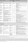
Table 1. Controlled clinical studies on the treatment of idiopathic sudden sensorineural hearing loss (systemic drug application)
Table 2
Table 2. Intratympanic drug application for the therapy of inner ear disorders (selection of clinical applications)
Table 3
Table 3. Conventional treatment of acute and chronic inner ear disorders in different countries
Table 4
Table 4. Grading scheme of outpatient and hospital-based tinnitus therapy (according to TRT-ADANO, from Wedel 2000 [219])
Figure 1
Figure 1. Ectopic, newly generated hair cells in the mammalian cochlea after Math1 gene transfer
Scanning electron microscopic images after in vivo inoculation of an adenovirus vector containing the Math1 gene (Ad.Math1.11D). The image reveals an ectopically located, regenerated hair cell in the area of the interdental cells of the limbus in the cochlea of the guinea pig. The hair cell features a well developed stereocilia bundle architecture. Even if hair cells should arise at micromechanically suboptimal points in the inner ear, this can lead to at least a partial restoration of hearing (see text). Scale bar: 2µm. From: Kawamoto et al. 2003 [118], © 2003 by the Society of Neuroscience, with kind permission of the author and publisher.
Figure 2
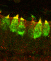
Figure 2. Generation of hair cells from embryonic stem-cells
Transplanted precursor cells of hair cells are integrated into the sensory epithelium and differentiate to become hair cells. The cylindrical hair cells (here in longitudinal section, and in green) feature bundles of sensory hairs (yellow, F-actin and epsin, superposed confocal images). From Li et al. 2003 [126], PNAS, © 2003 National Academy of Sciences, USA, with kind permission of the author and publishers.
Figure 3
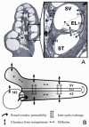
Figure 3. Principles of substance distribution in the inner ear
A: Cross section through the guinea pig cochlea and section of one turn. M: modiolus, ST: scala tympani, SV: scala vestibuli, EL: endolymphatic space. The "radial processes" considered in the 1D and 3D computer models take account of the exchange between the different compartments (scalae) and the clearance from the scalae /vestibulum into the body's blood circulation or into the modiolus. B: Depiction of "rolled up" cochlea and vestibulum: amongst the "longitudinal processes" include the geometric dimensions of the scalae, diffusion along the scalae, the helicotrema and into the vestibulum (V) and with local application to the middle ear (MO) the entry of substance through the round window membrane (RF). (after Plontke et al. 2004 and 2002, © Elsevier (a) and Otology & Neurotology Inc., with kind permission [177, 328]).
Figure 4
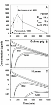
Figure 4. Glucocorticoid concentrations in the cochlea in animal experimental studies
A: Data from two similar animal-experimental studies imply large variations in the perilymph concentrations of locally applied glucocorticoids (Bachmann et al. 2001 and Parnes et al. 1999,[138, 160]). As such the clinical usefulness of these findings is very restricted. B: After considering the experimental differences between the two studies in the computer simulation, particularly regarding the artefacts from the sampling techniques, the pharmacokinetic profiles from the two studies could almost be brought to agreement (Plontke and Salt 2003,[183]). Consistent with model forecasts the concentrations in the basal turn in the guinea pig (B) appear to be similar to those in the basal turn of the human cochlea (C), since the diameters of the basal turn and the size of the round window membrane only differ a little. However, because of the longer length much lower concentrations are expected in the middle and apical turns in the human cochlea (C).
Figure 5
Figure 5. Location- and time-dependent methyl-prednisolone concentration in the scala tympani after intratympanic application
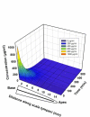
Calculation of methyl-prednisolone concentration in the scala tympani of the guinea pig cochlea after single application without volume stabilization reveals high peak concentrations at an early stage after the onset of application. The marked baso-apical concentration gradient is clearly recognizable. An even concentration distribution in the cochlea is first achieved only very late on and at a very low level (blue) compared to the peak concentration (red). The target structure in the cochlea, the relative vestibular-cochlear toxicity and the therapeutic range/index of the medication must therefore be considered especially with local application of medication to the round window membrane (data from Plontke and Salt 2003,[183]).
Figure 6
Figure 6. Baso-apical concentration gradient for different species
Since the longitudinal distribution of substances is determined essentially by diffusion and clearance, the concentration gradients depend on the length of the diffusion pathway, i.e. the length of the cochlear scalae. It can therefore be assumed that concentration gradients in larger cochlea are more extreme. This fact must be considered if the results from animal experiments involving different species are to be applied to the human situation. It is entirely conceivable that a substance applied to the round window membrane of the murine inner ear experiences no problems in gaining access to the apicocochlear area, while with the same application protocol in man effective medication levels may only be found in the basocochlear region.
Figure 7
Figure 7. Concentration gradients in the cochlea for two different application strategies
According to the exemplary calculations with a 3D computer model for substance distribution in the inner ear, short-term application for 30 min (A) shows clearly larger concentration gradients between basal and apical sections of the cochlea than does continuous drug application to the round window membrane in a saturated range (B). A concentration profile is also revealed in the cross-section that can not be registered using a 1D model. The numerical data in the legend displays the standardised concentrations. (from Plontke et al. 2004, © Elsevier) [328]
Figure 8
Figure 8. Acute hearing loss therapy by extracorporeal fibrinogen reduction
With H.E.L.P.® apheresis (heparin-induced extracorporeal lipoprotein fibrinogen precipitation, B.Braun, Medizintechnologie GmbH, Germany) LDL, lipoprotein (a) and fibrinogen is precipitated and filtered out at an acid pH of 5.12 in the extracorporeal plasma circulation using heparin. The filtered plasma then passes a heparin adsorber and a dialyzer to remove the buffer. The reduction in high-molecular-weight plasma proteins achieved this way improves the rheological properties of the blood.
Figure 9
Figure 9. Individual, tinnitus-associated activation maxima in the cortex
In order to achieve individual activation patterns associated with tinnitus perception in the cortex, PET images without tinnitus perception (suppression by i.v. lidocaine dosing) are subtracted from images obtained while tinnitus is being perceived. In this way it is possible to focus magnetic-stimulation on the individual, tinnitus associated activation maxima detected in the "on-off" PET studies (with kind permission of Dr. med. C. Plewnia and Dr. med. M. Reimold, Tübingen).
Figure 10
Figure 10. Neuronavigation guided transcranial magnet stimulation to modify the perception of tinnitus
Neuronavigation devices (e.g. BrainsightTM-Frameless, Magstim Co., Whitland, GB) allow the individual guidance of a TMS stimulation coil to a target identified by imaging (e.g. fMRT, PET, see fig. 10). In this way a targeted stimulation of predefined cortical areas is permitted (with kind permission of Dr C. Plewnia, Tübingen). A routine application of this procedure for tinnitus treatment is not justified on the basis of data obtained up until now.
Figure 11
Figure 11. Acute hearing loss guidelines of the DGHNO (2004): differential therapy of idiopathic sudden sensorineural hearing loss according to the audiogram type
Therapeutic recommendations for the differential treatment of acute, idiopathic impaired hearing according to the "acute hearing loss guideline" of the German Society for Otorhinolaryngology and Head & Neck Surgery (excerpts cited with the kind permission of the DGHNO) [266]. *With minor hearing loss the committee considers that an initial therapy with glucocorticoids alone over 3 days is feasible. If successful it should be continued circumstances permitting. Hyperbaric oxygenation is quoted in the guideline as a possible therapy for ISSNHL treated unsuccessfully by other means, and the guideline also recommends that the procedure be tested in prospective, randomised clinical studies.

References

    1. Forth W, Henschler D, Rummel W, Förstermann U, Starke K. Allgemeine und Spezielle Pharmakologie und Toxikologie. 8. Auflage ed. München, Jena: Urban & Fischer Verlag; 2001.
    1. Lamm K, Arnold W. How useful is corticosteroid treatment in cochlear disorders? Otorhinolaryngol Nova. 1999;9:203–216.
    1. Rarey KE, Luttge WG. Presence of type I and type II/IB receptors for adrenocorticosteroid hormones in the inner ear. Hear Res. 1989;41(2-3):217–221. - PubMed
    1. Rarey KE, Curtis LM, ten Cate WJ. Tissue specific levels of glucocorticoid receptor within the rat inner ear. Hear Res. 1993;64(2):205–210. - PubMed
    1. Rarey KE, Curtis LM. Receptors for glucocorticoids in the human inner ear. Otolaryngol Head Neck Surg. 1996;115(1):38–41. - PubMed

LinkOut - more resources