. 2009:8:Doc07.
doi: 10.3205/cto000059. Epub 2011 Mar 10.

Biomaterials in skull base surgery

Affiliations

Biomaterials in skull base surgery

Wolfgang Maier. GMS Curr Top Otorhinolaryngol Head Neck Surg. 2009.

Abstract

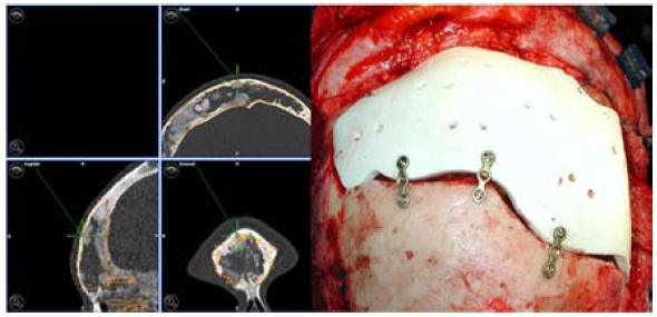
Reconstruction materials and techniques for the base of the skull have undergone rapid developments and differentiation in recent years. While mostly autotransplants, collagens or resorbable alloplastic materials are preferred for duraplasties, pronounced organ-specific differences can be observed in the reconstruction of hard tissues. The use of polymethylmethacryl bone cement, once wide-spread, has decreased greatly due to the release of toxic monomers. Bony autotransplants are still used primarily for smaller skull-base defects, intraoperatively formable titanium nets may be also used for larger fronto- or laterobasal reconstructions of bony defects. Defects in visible areas are increasingly closed with preformed titanium or ceramic implants, which are planned and fitted to the individual patient using preoperative CT imaging. At the skull base, this applies especially to reconstructions of the frontal sinus. For extensive reconstructions of the orbita, titanium nets and non-resorbable plastics have proven valuable; in closing smaller defects especially of the orbital floor, resorbable implants based on Polyglactin 901 are also used.

Keywords: computer assisted surgery; orbital reconstruction; prefabrication; skull base reconstruction.


Figures

Table 1
Table 1. Properties of Materials for Hard-tissue Reconstruction (modified from Eufinger et al. [22] and Peltola et al. [38])
Figure 1
Figure 1. Schematics of dura defect covering using the sandwich procedure. The alloplastic material is affixed with fibrin adhesive in an underlay procedure between the dura and bones and as a second layer overlaid on the bone. Additionally, nasal concha mucosa is placed over this (from Arndt et al. [8]).
Figure 2
Figure 2. Intraoperative situs before and after covering the dura defect. a: The left image shows exuding fluorescin (arrows) under blue-light filter, which was applied translumbar for localization diagnostics of the defect. b: In the right image, the defect has already been closed in the sandwich procedure (arrow: Ethisorb®); the conchal mucosa has not yet been applied (modified and supplemented from [8]).
Figure 3
Figure 3. Sagittal CT of a paranasal sinus carcinoma with destruction of the rhinobase, dura infiltration and elevation of the frontal brain.
Figure 4
Figure 4. Intraoperative situs of the patient in Fig. 3 after interdisciplinary resection (ORL/Neurosurgery) of the tumor including the dura via a transfrontal-transbasal access. a: Left: titanium mesh in situ (arrows), fixed with titanium miniscrews. The frontal brain is lifted with the spatula. b: Right: collagen fleece (arrows) as second layer on the titanium mesh; before the frontal brain is tipped back and before the dura is closed using galeaperiostium.
Figure 5
Figure 5. T2-weighted MRT in coronar projection of a woman with status after temporobasal giant-cell granuloma, Status after dura resection and duraplasty and reconstruction of the bony laterobase using titanium mesh (arrows). The titanium mesh is recognized in the extinction in the MRT and effects further extinction artifacts in the immediate vicinity.
Figure 6
Figure 6. Prefabrication of hard-tissue replacement in a woman with frontal, frontobasal and ethmoidal fibrous dysplasia.
a: Left: preoperative planning surgery and model: the planned resection zones corresponding to the volume of the implant to be preformed are marked red in the CT-dataset (from Dämmrich et al. [41]). b: Right: intraoperative image of the computer-assisted operation: the navigation pointer (green) points to the resection boundaries which were determined in the preoperative planning surgery (orange).
Figure 7
Figure 7. Patient from Fig. 6 after fitting of the preformed implant. a: Left: The navigation pointer (green) shows the exact agreement of the surfaces of implant and preoperative bone (orange). b: Right: ceramic implant (Bioverit II®) in situ, fixed with titanium plates (from [40]).

Similar articles

Cited by

References

    1. Potter JK, Ellis E. Biomaterials for the reconstruction of the internal orbit. J Oral Maxillofac Surg. 2004;62:1280–1297. doi: 10.1016/j.joms.2004.04.018. Available from: http://dx.doi.org/10.1016/j.joms.2004.04.018. - DOI - DOI - PubMed
    1. Castelnouvo PG, Delu G, Locatelli D, Padoan G, de Bernardi F, Pistochini A, Bignami M. Endonasal endoscopic duraplasty: Our experience. Skull Base. 2006;16:19–23. doi: 10.1055/s-2005-922096. Available from: http://dx.doi.org/10.1055/s-2005-922096. - DOI - DOI - PMC - PubMed
    1. Aletsee C, Konopik V, Dazert S, Dieler R. Operative Versorgung von Verletzungen der Rhinobasis. Laryngo-Rhino-Otol. 2003;82:626–631. doi: 10.1055/s-2003-42688. Available from: http://dx.doi.org/10.1055/s-2003-42688. - DOI - DOI - PubMed
    1. Draf W, Schick B. How I do it: Endoscopic-microscopic anterior skull base reconstruction. Skull Base. 2007;17:53–58. doi: 10.1055/s-2006-959335. Available from: http://dx.doi.org/10.1055/s-2006-959335. - DOI - DOI - PMC - PubMed
    1. Schick B, Ibing R, Brors D, Draf W. Longterm study in endonasal duraplasty and review of the literature. Ann Otol Laryngol Rhinol. 2001;114:142–147. - PubMed