. 2009:8:Doc10.
doi: 10.3205/cto000062. Epub 2011 Mar 10.

Biomaterials in cochlear implants

Affiliations

Biomaterials in cochlear implants

Timo Stöver et al. GMS Curr Top Otorhinolaryngol Head Neck Surg. 2009.

Abstract

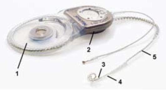
The cochlear implant (CI) represents, for almost 25 years now, the gold standard in the treatment of children born deaf and for postlingually deafened adults. These devices thus constitute the greatest success story in the field of 'neurobionic' prostheses. Their (now routine) fitting in adults, and especially in young children and even babies, places exacting demands on these implants, particularly with regard to the biocompatibility of a CI's surface components. Furthermore, certain parts of the implant face considerable mechanical challenges, such as the need for the electrode array to be flexible and resistant to breakage, and for the implant casing to be able to withstand external forces.As these implants are in the immediate vicinity of the middle-ear mucosa and of the junction to the perilymph of the cochlea, the risk exists - at least in principle - that bacteria may spread along the electrode array into the cochlea. The wide-ranging requirements made of the CI in terms of biocompatibility and the electrode mechanism mean that there is still further scope - despite the fact that CIs are already technically highly sophisticated - for ongoing improvements to the properties of these implants and their constituent materials, thus enhancing the effectiveness of these devices.This paper will therefore discuss fundamental material aspects of CIs as well as the potential for their future development.

Keywords: biocompatibility; biomaterials; coating; cochlear implant; cochleostomy; drug delivery; electrode; inner ear; nanoparticles; surface functionalization.


Figures

Figure 1
Figure 1. The implantable portion of the cochlear implant: (1) Transmitter coil in a silicone sheath; (2): Electronics enclosed in a titanium casing; (3): Electrode with platinum contacts in a silicone array; (4): Cochleostomy site; (5): Silicone cable in the middle-ear/mastoid region.
Figure 2
Figure 2. Preoperative marking of the incision line for the “Hannover C” incision formerly used, showing the large wound surface that results.
Figure 3
Figure 3. Smaller, retroauricular incision in use today (left ear, dummy implant).
Figure 4
Figure 4. View of preformed electrode with modified tip (Softip®, Cochlear Ltd).
Figure 5
Figure 5. Histological section through the cochlea of a guinea pig showing formation of new connective tissue and bone in the area of the scala tympani (EK: electrode channel; scala vestibuli (Sc. V.); scala media (Sc. M.); RK: Rosenthal channel, BDG: connective tissue, K: bone).
Figure 6
Figure 6. View of the cochleostomy during the insertion of a cochlear implant electrode.
Figure 7
Figure 7. Cochlear implant with cracked casing (here, ceramic casing) following application of mechanical force caused by the wearer falling on the implant.
Figure 8
Figure 8. Individual compatibility test designed to rule out allergic reactions to the cochlear implant material. Left: preparation of the forearm; centre: incision for the creation of a subcutaneous pocket; right: positioning of the test material beneath the skin.
Figure 9
Figure 9. Swelling around the implant site in a case where the implant is chronically infected. Right of picture: spontaneous perforation of the skin.
Figure 10
Figure 10. Infected cochlear implant with marked formation of granulation tissue around the implant.
Figure 11
Figure 11. Section through a temporal bone and the cochlea with inserted perimodiolar cochlear implant electrodes in the scala tympani, showing the electrode contact surfaces facing towards the modiolus.
Figure 12
Figure 12. The development of biomimetic implants: different methods of surface functionalization.
Figure 13
Figure 13. Schematic representation of physical surface functionalization: alteration of surface topography.
Figure 14
Figure 14. Example of linear microstructure on silicone, generated through ablation by ultrashort laser pulses.
Figure 15
Figure 15. Example of physical surface functionalization: cultivation of cells growing adherently (fibroblasts, duration of culture: three days) on laser-structured silicone surface (structural width 10 µm).
Figure 16
Figure 16. Schematic representation of biochemical surface functionalization: binding of signal molecules onto the electrode.
Figure 17
Figure 17. Schematic representation of chemical surface functionalization: binding of polymer chains onto the electrode.
Figure 18
Figure 18. Schematic representation of biological surface functionalization: binding of adherent cells onto the electrode.
Figure 19
Figure 19. A representative example showing the consequences of deafness in the region of the spiral ganglion. Histological view of the spiral ganglion cells (SGC) in the Rosenthal channel (A) when hearing was normal, (B) six weeks after deafening, with most of the SGCs having degenerated. Sc.T. scala tympani.
Figure 20
Figure 20. Schematic representation of drug delivery from the electrode body. Use of the cochlear implant as a means of access for local drug delivery.
Figure 21
Figure 21. Model investigation of electrode prototypes for delivery of fluids within the cochlea. Shown here: release of a dye at both the tip and side of the electrode array as a means of intracochlear fluid application.
Figure 22
Figure 22. Schematic structure of a cochlear implant with integral micropump (MedEl GmbH) showing a septum port between the implantable pump (i.e. the circular structure on the right) and the implant (left).

Similar articles

Cited by

References

    1. Burgio P. Safety considerations of cochlear implantation. Otolaryngol Clin North Am. 1986;19(2):237–247. - PubMed
    1. Lehnhardt E. Biokompatibilität der Cochlear-implants. Eur Arch Otorhinolaryngol Suppl. 1992;1:223–233. - PubMed
    1. BQS Qualitätsreport. Düsseldorf: BQS Institut für Qualität und Patientensicherheit; 2007. p. 36. Available from: http://www.bqs-qualitaetsreport.de/
    1. Dunn CC, Tyler RS, Oakley S, Gantz BJ, Noble W. Comparison of speech recognition and localization performance in bilateral and unilateral cochlear implant users matched on duration of deafness and age at implantation. Ear Hear. 2008;29(3):352–359. doi: 10.1097/AUD.0b013e318167b870. Available from: http://dx.doi.org/10.1097/AUD.0b013e318167b870. - DOI - DOI - PMC - PubMed
    1. O'Donoghue GM, Nikolopoulos TP. Minimal access surgery for pediatric cochlear implantation. Otol Neurotol. 2002;23(6):891–894. doi: 10.1097/00129492-200211000-00014. Available from: http://dx.doi.org/10.1097/00129492-200211000-00014. - DOI - DOI - PubMed