

Nasal surgery in patients with systemic disorders
- PMID: 22073106
- PMCID: PMC3199829
- DOI: 10.3205/cto000066
Nasal surgery in patients with systemic disorders
Abstract
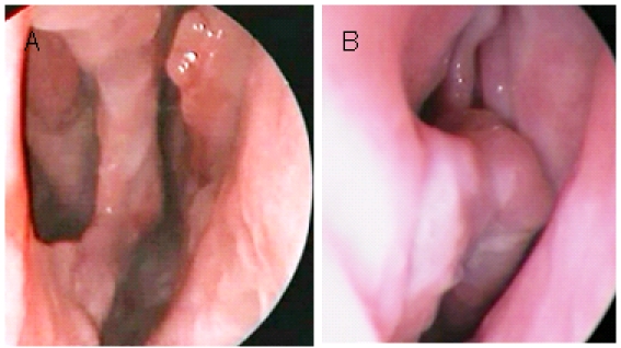
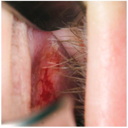
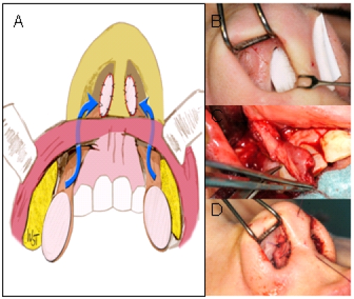
Multisystemic disorders represent a heterogenous group of diseases which can primarily manifest at the nose and paranasal sinuses as limited disease or secondarily as part of systemic involvement. Rhinologists therefore play an important role in the diagnostic but also therapeutic process. Although therapy of multisystemic disorders is primary systemic, additional rhinosurgery may become necessary. The spectrum of procedures consists of sinus surgery, surgery of the orbit and lacrimal duct, septorhinoplasty and closure of nasal septal perforation. Since the prevalence of most systemic diseases is very rare, recommendations are based on the analysis of single case reports and case series with a limited number of patients only. Although data is still limited, experiences published so far have shown that autologous cartilage or bone grafts can be used in nasal reconstruction of deformities caused by tuberculosis, leprosy, Wegener's granulomatosis, sarcoidosis and relapsing polychondritis. Experiences gained from these diseases support the concept that well-established techniques of septorhinoplasty can be used in systemic diseases as well. However, a state of remission is an essential condition before considering any rhinosurgery in these patients. Even under these circumstances revision surgery has to be expected more frequently compared to the typical collective of patients undergoing septorhinoplasty. In addition, experiences gained from saddle nose reconstruction may in part be of value for the treatment of nasal septal perforations since implantation of cartilage grafts often represents an essential step in multilayer techniques of closure of nasal septal perforations. Aside from the treatment of orbital complications sinus surgery has been proven beneficial in reducing nasal symptoms and increasing quality of life in patients refractory to systemic treatment.
Keywords: nasal septal perforation; rhinoplasty; saddle nose; sinusitis; systemic disorder.
Figures
References
-
- Hup AK, Haitjema T, de Kuijper G. Primary nasal tuberculosis. Rhinology. 2001;39(1):47–48. - PubMed
-
- Prasad KC, Sreedharan S, Chakravarthy Y, Prasad SC. Tuberculosis in the head and neck: experience in India. J Laryngol Otol. 2007;121(10):979–985. doi: 10.1017/S0022215107006913. Available from: http://dx.doi.org/10.1017/S0022215107006913. - DOI - DOI - PubMed
-
- Kim YM, Kim AY, Park YH, Kim DH, Rha KS. Eight cases of nasal tuberculosis. Otolaryngol Head Neck Surg. 2007;137(3):500–504. doi: 10.1016/j.otohns.2007.04.009. Available from: http://dx.doi.org/10.1016/j.otohns.2007.04.009. - DOI - DOI - PubMed
-
- Choi YC, Park YS, Jeon EJ, Song SH. The disappeared disease: tuberculosis of the nasal septum. Rhinology. 2000;38(2):90–92. - PubMed
-
- Lai TY, Liu PJ, Chan LP. Primary nasal tuberculosis presenting with septal perforation. J Formos Med Assoc. 2007;106(11):953–955. doi: 10.1016/S0929-6646(08)60066-2. Available from: http://dx.doi.org/10.1016/S0929-6646(08)60066-2. - DOI - DOI - PubMed
LinkOut - more resources
Full Text Sources
