

Quantitative evaluation of scintillation camera imaging characteristics of isotopes used in liver radioembolization
- PMID: 22073149
- PMCID: PMC3207835
- DOI: 10.1371/journal.pone.0026174
Quantitative evaluation of scintillation camera imaging characteristics of isotopes used in liver radioembolization
Abstract
Background: Scintillation camera imaging is used for treatment planning and post-treatment dosimetry in liver radioembolization (RE). In yttrium-90 (90Y) RE, scintigraphic images of technetium-99m (99mTc) are used for treatment planning, while 90Y Bremsstrahlung images are used for post-treatment dosimetry. In holmium-166 (166Ho) RE, scintigraphic images of 166Ho can be used for both treatment planning and post-treatment dosimetry. The aim of this study is to quantitatively evaluate and compare the imaging characteristics of these three isotopes, in order that imaging protocols can be optimized and RE studies with varying isotopes can be compared.
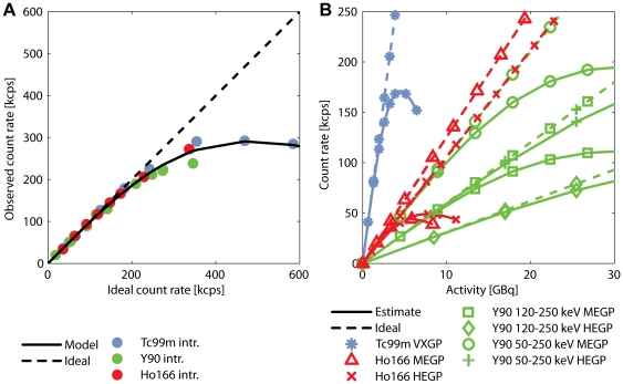
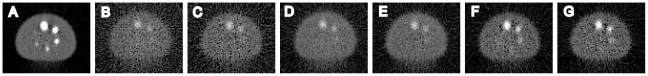
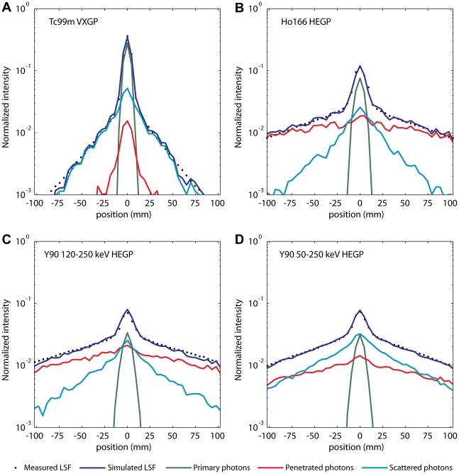
Methodology/principal findings: Phantom experiments were performed in line with NEMA guidelines to assess the spatial resolution, sensitivity, count rate linearity, and contrast recovery of 99mTc, 90Y and 166Ho. In addition, Monte Carlo simulations were performed to obtain detailed information about the history of detected photons. The results showed that the use of a broad energy window and the high-energy collimator gave optimal combination of sensitivity, spatial resolution, and primary photon fraction for 90Y Bremsstrahlung imaging, although differences with the medium-energy collimator were small. For 166Ho, the high-energy collimator also slightly outperformed the medium-energy collimator. In comparison with 99mTc, the image quality of both 90Y and 166Ho is degraded by a lower spatial resolution, a lower sensitivity, and larger scatter and collimator penetration fractions.
Conclusions/significance: The quantitative evaluation of the scintillation camera characteristics presented in this study helps to optimize acquisition parameters and supports future analysis of clinical comparisons between RE studies.
Conflict of interest statement
Figures


References
-
- Kennedy AS, Salem R. Radioembolization (yttrium-90 microspheres) for primary and metastatic hepatic malignancies. Cancer J. 2010;16:163–175. - PubMed
-
- Vente MA, Wondergem M, van der Tweel I, van den Bosch MA, Zonnenberg BA, et al. Yttrium-90 microsphere radioembolization for the treatment of liver malignancies: a structured meta-analysis. Eur Radiol. 2009;19:951–959. - PubMed
-
- Gulec SA, Fong Y. Yttrium 90 microsphere selective internal radiation treatment of hepatic colorectal metastases. Arch Surg. 2007;142:675–682. - PubMed
-
- Fabbri C, Sarti G, Cremonesi M, Ferrari M, Di DA, et al. Quantitative analysis of 90Y Bremsstrahlung SPECT-CT images for application to 3D patient-specific dosimetry. Cancer Biother Radiopharm. 2009;24:145–154. - PubMed
-
- Minarik D, Sjogreen GK, Ljungberg M. Evaluation of quantitative (90)Y SPECT based on experimental phantom studies. Phys Med Biol. 2008;53:5689–5703. - PubMed
Publication types
MeSH terms
Substances
LinkOut - more resources
Full Text Sources
Other Literature Sources
