. 2011;6(11):e27079.
doi: 10.1371/journal.pone.0027079. Epub 2011 Nov 2.

Neural stem/progenitor cells from the adult human spinal cord are multipotent and self-renewing and differentiate after transplantation

Affiliations

Neural stem/progenitor cells from the adult human spinal cord are multipotent and self-renewing and differentiate after transplantation

Andrea J Mothe et al. PLoS One. 2011.

Abstract

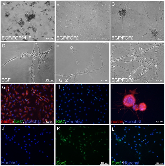
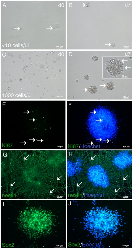
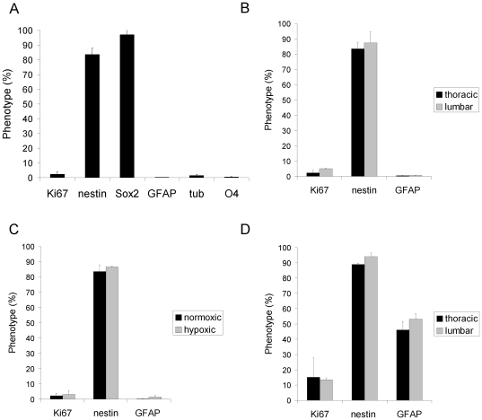
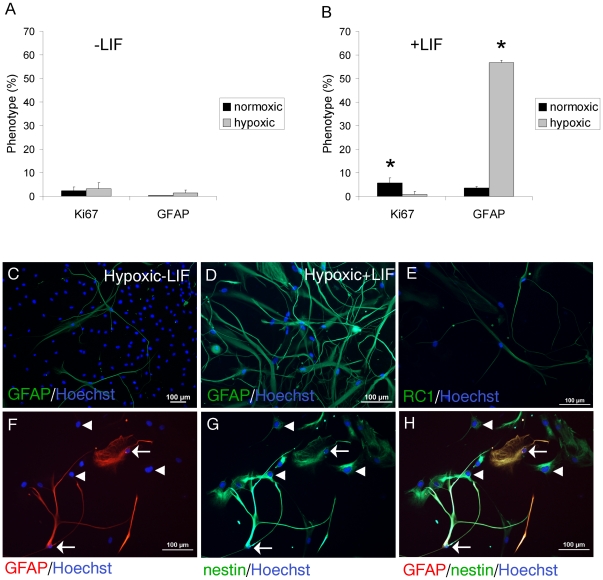
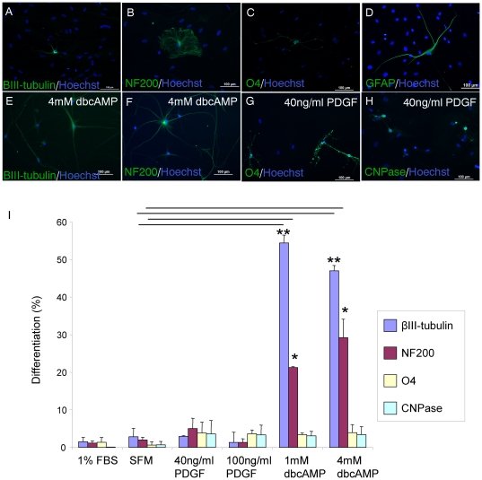
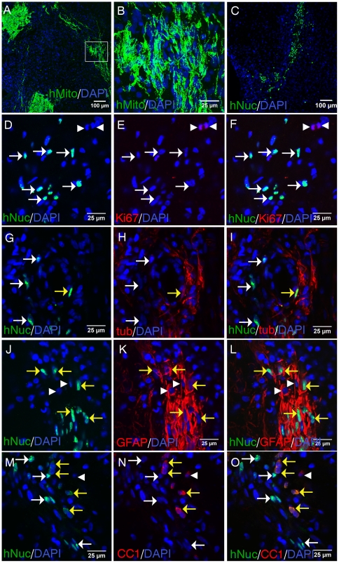
Neural stem/progenitor cell (NSPC) transplantation is a promising therapy for spinal cord injury (SCI). However, little is known about NSPC from the adult human spinal cord as a donor source. We demonstrate for the first time that multipotent and self-renewing NSPC can be cultured, passaged and transplanted from the adult human spinal cord of organ transplant donors. Adult human spinal cord NSPC require an adherent substrate for selection and expansion in EGF (epidermal growth factor) and FGF2 (fibroblast growth factor) enriched medium. NSPC as an adherent monolayer can be passaged for at least 9 months and form neurospheres when plated in suspension culture. In EGF/FGF2 culture, NSPC proliferate and primarily express nestin and Sox2, and low levels of markers for differentiating cells. Leukemia inhibitory factor (LIF) promotes NSPC proliferation and significantly enhances GFAP expression in hypoxia. In differentiating conditions in the presence of serum, these NSPC show multipotentiality, expressing markers of neurons, astrocytes, and oligodendrocytes. Dibutyryl cyclic AMP (dbcAMP) significantly enhances neuronal differentiation. We transplanted the multipotent NSPC into SCI rats and show that the xenografts survive, are post-mitotic, and retain the capacity to differentiate into neurons and glia.Together, these findings reveal that multipotent self-renewing NSPC cultured and passaged from adult human spinal cords of organ transplant donors, respond to exogenous factors that promote selective differentiation, and survive and differentiate after transplantation into the injured spinal cord.


Conflict of interest statement

Competing Interests: The authors have declared that no competing interests exist.

Figures

Figure 1
Figure 1. Adult human spinal cord NSPC require an adherent substrate for selection and expansion in EGF/FGF2 culture.
A, NSPC were cultured from the time of isolation in uncoated tissue culture flasks. Phase contrast image shows primarily aggregates of debris and some cells at 43div in hypoxic EGF/FGF2 culture containing LIF. B-C, In contrast, in the presence of an adhesive substrate, NSPC are selected in EGF/FGF2 medium and can be expanded continuously. B-C, NSPC cultured and passaged as a monolayer on matrigel for 41div (B) can also form neurospheres in adherent culture (C). D-F, NSPC were cultured for 48div as an adherent monolayer on matrigel. In media containing only EGF (D) or FGF2 (E) fewer cells survived compared to media containing both EGF and FGF2 (F). Both EGF and FGF2 were required for expansion and neurosphere formation. Almost all NSPC expressed nestin (red) (G, 26div). NSPC also expressed Ki67 proliferating nuclear antigen (G, green in overlay; H, green; blue, Hoechst nuclear counterstain). I, Cluster of neurospheres were removed from adherent culture (25div), plated on matrigel coated well in EGF/FGF2 medium, and stained with nestin. Most NSPC expressed Sox2 (66div shown) (K, green; J, corresponding Hoechst staining of nuclei; L, merged).
Figure 2
Figure 2. Neurospheres form at clonal density and express stem cell markers.
NPSC cultured as an adherent monolayer for 39div were seeded at clonal density (<10 cells/µl) in EGF/FGF2 medium in suspension culture. A, Image taken on the day of seeding (d0) show single cells (arrows). B, 7d later neurospheres have formed (arrows). C, NSPC seeded at higher density (1000 cells/µl) at the time of plating form more neurospheres which are larger (arrows) (D, at 7d). Inset in D shows a high magnification image of a single neurosphere with a phase bright profile and ciliary projections (arrows). Neurospheres were plated onto matrigel and immunostained with stem cell markers. E-F, A single neurosphere showing proliferating Ki67+ NSPC (arrows) (E, green) stained with the nuclear dye Hoechst (F, merged). G-H, Three adjacent neurospheres expressing high levels of nestin (G, green) with nestin+ processes radially emanating from the neurospheres (arrows) (H, merged). I-J, Most NSPC comprising the neurosphere express Sox2 (I, green; J, merged with Hoechst nuclear counterstain).
Figure 3
Figure 3. Phenotypic expression profile of human spinal cord NSPC in EGF/FGF2 culture.
A, NPSC primarily express nestin and Sox2, and low levels of GFAP, tubulin, and O4. Data shown is averaged from cultured cells generated from the thoracic cord of 3 independent cultures at 35, 66, and 83div. B, NSPC generated from the thoracic and lumbar spinal cord from 2 cultures (34, 66, and 82div) show similar expression levels. C, NSPC cultured in normoxic or hypoxic conditions (2 cultures at 45 and 58div) show no significant differences in Ki67, nestin, and GFAP expression. D, NSPC derived from a younger donor (34 and 45div) also show similar expression levels when cells were generated from the thoracic or lumbar spinal cord. Cultures from the young donor (D) showed significantly higher GFAP expression than cultures from the older donors (B) (p = 0.007).
Figure 4
Figure 4. LIF enhances GFAP expression of adult human spinal cord NSPC in EGF/FGF2 culture.
A, In the absence of LIF (3 cultures at 42, 55 and 66div), there is no significant difference in Ki67 or GFAP expression in normoxic or hypoxic conditions. B, In the presence of LIF (3 cultures at 35, 55, and 66div), there is a significant increase in Ki67 expression in normoxic versus hypoxic conditions (* p<0.025). LIF significantly increases GFAP expression in hypoxia (** p<0.001). C, GFAP+ NSPC in hypoxic culture in the absence of LIF. D, There are more GFAP+ cells in hypoxia + LIF. E, RC1+ NSPC (39div) in normoxic conditions. F, GFAP+ cells (arrows) are double-labelled with nestin (G) as shown in the merged panel (H). Arrows show GFAP+/nestin+cells. Arrowheads show GFAP-/nestin+cells.
Figure 5
Figure 5. Adult human spinal cord NSPC are multipotent and directed differentiation can be promoted with exogenous factors.
A-D, NPSC (from 3 cultures at 46, 58, and 72div) were plated in 1% FBS for 4wk and immunostained with cell type specific markers. NSPC expressed markers of neurons, βIII-tubulin (A) and NF200 (B), oligodendrocyte progenitor cell marker O4 (C), and GFAP (D) for astrocytes. The addition of dbcAMP increased the expression of βIII-tubulin (E) and NF200 (F). PDGF increased O4 (G) and CNPase (H) expression. I, Quantitation of % phenotype of NSPC after 4wk in dbcAMP, PDGF, and FBS or SFM controls. The expression of βIII-tubulin significantly increased in the presence of dbcAMP relative to SFM controls (** p<0.001). NF200 expression was also significantly increased compared to controls (* p<0.004). There was no significant difference in the quantitative expression of O4 and CNPase in any of the groups.
Figure 6
Figure 6. Transplantation of adult human spinal cord NSPC into the injured rat spinal cord.
Adult human spinal cord NSPC were transplanted rostral and caudal to the lesion site at 1wk post-SCI. Rats were sacrificed at 1wk post-transplantation and tissue was sectioned in a parasagittal orientation. A, Low magnification fluorescent image showing transplanted cells (green) identified with human specific mitochondrial antigen (hMito). Tissue was stained with DAPI nuclear counterstain (blue). B, High magnification confocal image of boxed area showing hMito+ transplanted cells. C, Low magnification image showing transplanted cells (green) identified with human specific nuclear antigen (hNuc). D-F, Ki67+ (red) proliferating cells (arrowheads) adjacent to hNuc+ (green) transplanted cells. G-I, Transplanted NSPC expressing βIII-tubulin (tub); hNuc+/tub+ (yellow arrows); hNuc+/tub- cells (white arrows). J-L, Transplanted NSPC expressing GFAP; hNuc+/GFAP+ (yellow arrows); hNuc-/GFAP+ (arrowheads). M-O, Transplanted NSPC expressing CC1; hNuc+/CC1+ (yellow arrows); hNuc+/CC1- (white arrows); hNuc-/CC1+ (arrowheads).

References

    1. Kwon BK, Sekhon LH, Fehlings MG. Emerging repair, regeneration, and translational research advances for spinal cord injury. Spine (Phila Pa 1976) 2010;35:S263–270. - PubMed
    1. Tator CH. Review of treatment trials in human spinal cord injury: issues, difficulties, and recommendations. Neurosurgery. 2006;59:957–982; discussion 982-957. - PubMed
    1. Reynolds BA, Weiss S. Generation of neurons and astrocytes from isolated cells of the adult mammalian central nervous system. Science. 1992;255:1707–1710. - PubMed
    1. Gage FH. Mammalian neural stem cells. Science. 2000;287:1433–1438. - PubMed
    1. Bjorklund LM, Sanchez-Pernaute R, Chung S, Andersson T, Chen IY, et al. Embryonic stem cells develop into functional dopaminergic neurons after transplantation in a Parkinson rat model. Proc Natl Acad Sci U S A. 2002;99:2344–2349. - PMC - PubMed

Publication types