Clinical Trial
. 2011;55(3):e31.
doi: 10.4081/ejh.2011.e31. Epub 2011 Aug 27.

Lubricin is expressed in chondrocytes derived from osteoarthritic cartilage encapsulated in poly (ethylene glycol) diacrylate scaffold

Affiliations
Clinical Trial

Lubricin is expressed in chondrocytes derived from osteoarthritic cartilage encapsulated in poly (ethylene glycol) diacrylate scaffold

G Musumeci et al. Eur J Histochem. 2011.

Abstract

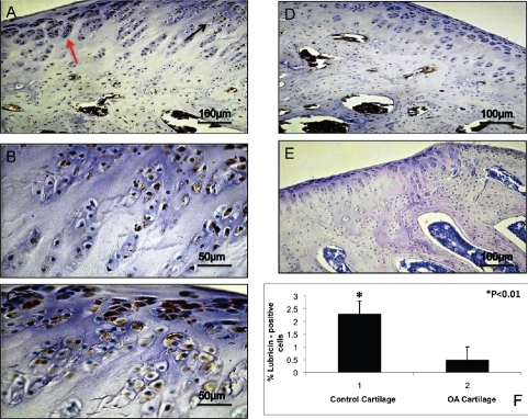
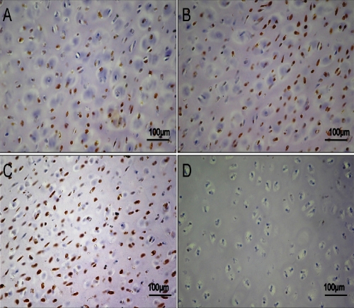
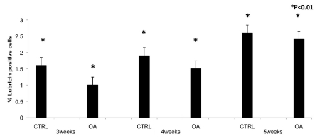
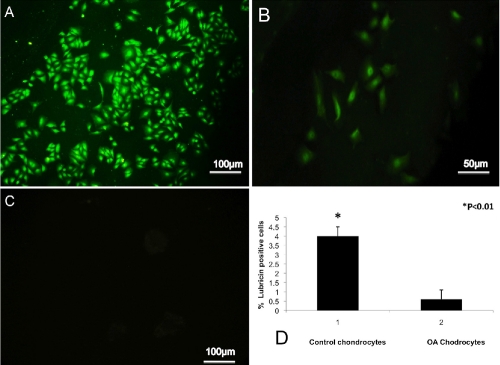
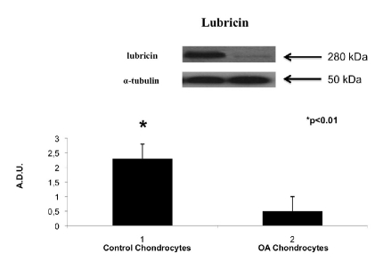
Osteoarthritis (OA) is characterized by degenerative changes within joints that involved quantitative and/or qualitative alterations of cartilage and synovial fluid lubricin, a mucinous glycoprotein secreted by synovial fibroblasts and chondrocytes. Modern therapeutic methods, including tissue-engineering techniques, have been used to treat mechanical damage of the articular cartilage but to date there is no specific and effective treatment. This study aimed at investigating lubricin immunohistochemical expression in cartilage explant from normal and OA patients and in cartilage constructions formed by Poly (ethylene glycol) (PEG) based hydrogels (PEG-DA) encapsulated OA chondrocytes. The expression levels of lubricin were studied by immunohistochemistry: i) in tissue explanted from OA and normal human cartilage; ii) in chondrocytes encapsulated in hydrogel PEGDA from OA and normal human cartilage. Moreover, immunocytochemical and western blot analysis were performed in monolayer cells from OA and normal cartilage. The results showed an increased expression of lubricin in explanted tissue and in monolayer cells from normal cartilage, and a decreased expression of lubricin in OA cartilage. The chondrocytes from OA cartilage after 5 weeks of culture in hydrogels (PEGDA) showed an increased expression of lubricin compared with the control cartilage. The present study demonstrated that OA chondrocytes encapsulated in PEGDA, grown in the scaffold and were able to restore lubricin biosynthesis. Thus our results suggest the possibility of applying autologous cell transplantation in conjunction with scaffold materials for repairing cartilage lesions in patients with OA to reduce at least the progression of the disease.

Keywords: chondrocytes; hydrogels (PEG-DA); lubricin.; osteoarthritis.


Figures

Figure 1
Figure 1
A) Lubricin immunohistochemistry (cytoplasmic and perinuclear immunola-beling) specimen from control cartilage (tissue explanted); strong lubricin immunolabeling of chondrocytes; magnification ×20; scale bar: 100 µm. B) Magnification of panel A, black arrow; magnification ×40; scale bar: 50 µm. C) magnification of panel A, red arrow; magnification ×40; scale bar: 50 µm. D) Lubricin immunohistochemistry specimen from OA cartilage (tissue explanted); weak lubricin immunolabeling of chondrocytes; magnification ×20; scale bar: 100 µm. E) No immunoreaction was observed in the negative control treated with PBS without the primary antibodies (tissue explanted); magnification ×20; scale bar: 100 µm. F) Percentage of lubricin positive cells out of the total number of cells counted in control cartilage and in OA cartilage.
Figure 2
Figure 2
A) Lubricin immunohistochemistry (cytoplasmic and perinuclear immunola-beling) specimen from control chondrocytes after 3 weeks of encapsulations in PEGDA hydrogels. Strong lubricin immunolabeling in chondrocytes; magnification ×20; scale bar: 100 µm. B) Lubricin immunostained specimen from control chondrocytes after 4 weeks of encapsulation in PEGDA hydrogels. Strong lubricin immunolabeling in chondrocytes; magnification ×20; scale bar: 100 µm. C) Lubricin immunostained specimen from control chondrocytes after 5 weeks of encapsulations in PEGDA hydrogels; very strong lubricin immunolabeling in chondrocytes; magnification ×20; scale bars: 100 µm. D) No immunoreaction was observed in the negative control treated with PBS without the primary antibodies; magnification ×20; scale bars: 100 µm.
Figure 3
Figure 3
A) Lubricin immunohistochemistry specimen from OA chondrocytes after 3 weeks of encapsulations in PEGDA hydrogels; weak lubricin immunolabeling in chondrocytes; magnification ×20; scale bars: 100 µm. B) Magnification (×40) of the panel A; scale bar: 50 µm. C) Lubricin immunostained specimen from OA chondrocytes after 4 weeks of encapsulation in PEGDA hydrogels; strong lubricin immunolabeling in the chondrocytes; magnification ×20; scale bars: 100 µm. D) Lubricin immunostained specimen from OA chondrocytes after 5 weeks of encapsulation in PEGDA hydrogels; very strong lubricin immunolabeling in the chondrocytes; magnification ×20; scale bar: 100 µm.
Figure 4
Figure 4
Percentage of lubricin positive cells out of the total number of cells counted in control chondrocytes and in OA chondrocytes after 3, 4 and 5 weeks in PEGDA hydrogels.
Figure 5
Figure 5
A) Lubricin immunocytochemistry (cytoplasmic and perinuclear immunolabeling) specimen from control chondrocytes; strong lubricin immunolabeling in cells at fourth passage of culture; magnification ×20; scale bars: 100 µm. B) Lubricin immunocytochemistry (cytoplasmic and perinuclear immunolabeling) specimen from OA chondrocytes; weak lubricin immunolabeling in cells at fourth passage of culture; magnification ×20; scale bar: 50 µm. C) No immunoreaction was observed in the negative control treated with PBS without the primary antibodies; magnification ×20; scale bar: 100 µm. D) Percentage of lubricin positive cells out of the total number of cells counted in control chondrocytes and in OA chondrocytes.
Figure 6
Figure 6
Lubricin expression induced in human monolayer chondrocytes from normal cartilage and human monolayer chondrocytes from OA cartilage determined by Western blot analysis. Data show the relative expression (mean ± SEM) of lubricin calculated as arbitrary densitometric units (A.D.U.) collected from three independent experiments. *P<0.01 compared to monolayer chondrocytes.

References

    1. Loreto C, Musumeci G, Leonardi R. Chondrocyte-like apoptosis in temporomandibular joint disc internal derangement as a repair-limiting mechanism. An in vivo study. Histol Histopathol. 2009;24:293–8. - PubMed
    1. Musumeci G, Loreto C, Carnazza ML, Martinez G. Characterization of apoptosis in articular cartilage derived from the knee joints of patients with osteoarthritis. Knee Surg Sports Traumatol Arthrosc. 2011;19:307–13. - PubMed
    1. Schulze-Tanzil G. Activation and dedifferentiation of chondrocytes: implications in cartilage injury and repair. Ann Anat. 2009;191:325–38. - PubMed
    1. Burrage PS, Mix KS, Brinckerhoff CE. Matrix metalloproteinases: role in arthritis. Front Biosci. 2006;11:529–43. - PubMed
    1. Pritzker KP, Gay S, Jimenez SA, Ostergaard K, Pelletier JP, Revell PA, et al. Osteoarthritis cartilage histopathology: grading and staging. Osteoarthritis Cartilage. 2006;14:13–29. - PubMed

Publication types

MeSH terms