Review
. 2012 Jan 13;287(3):1612-9.
doi: 10.1074/jbc.R111.301150. Epub 2011 Nov 10.

Chemistry and biology of vision

Affiliations
Review

Chemistry and biology of vision

Krzysztof Palczewski. J Biol Chem. .

Abstract

Visual perception in humans occurs through absorption of electromagnetic radiation from 400 to 780 nm by photoreceptors in the retina. A photon of visible light carries a sufficient amount of energy to cause, when absorbed, a cis,trans-geometric isomerization of the 11-cis-retinal chromophore, a vitamin A derivative bound to rhodopsin and cone opsins of retinal photoreceptors. The unique biochemistry of these complexes allows us to reliably and reproducibly collect continuous visual information about our environment. Moreover, other nonconventional retinal opsins such as the circadian rhythm regulator melanopsin also initiate light-activated signaling based on similar photochemistry.


Figures

FIGURE 1.
FIGURE 1.
Structures of rod and cone OS and ROS internal membranes. A, neuronal organization of a typical mammalian retina. A cross-sectional representation of rod and cone photoreceptors is presented, illustrating their connections to the RPE distally and to relaying cells (bipolar, horizontal, amacrine, and ganglion cells) proximally. The rod structure has a longer OS with membrane-enclosed disks tightly packed without connections to the plasma membrane. Cone disks are continuously connected with the plasma membrane. This figure was reprinted from Ref. with permission. B, electron tomogram of vitrified ROS. The electron tomogram is represented in three orthogonal slices through the ROS volume. An x-y slice (right) and a y-z slice (left) display the high order and regular arrangement of stacked disks. Red represents the high concentrations of rhodopsin found in disk membranes; spacer structures (pillars) are colored green. Scale bar = 200 nm. C, blueprint of ROS. A schematic of a plasma membrane and two disks with measured distances between membrane components is shown. Green cylinders represent monomeric rhodopsin, which forms a larger cluster in native ROS. B and C were reprinted from Ref. with permission.
FIGURE 2.
FIGURE 2.
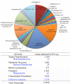
Transcriptome analysis of wild-type mouse eye. RNA sequencing of WT mouse eye reveals the transcriptional landscape of this tissue and the precise quantification of transcripts present. A breakdown of assigned transcripts is presented along with the number of transcripts in each category. The table highlights key Gene Ontology (GO) term categories and subcategories that relate to different aspects of visual processing. Notable are 2570 transcripts of unknown function of a total of 13,406 transcripts detected in WT eye, prospects for new avenues of vision research. Data shown are reprinted from Ref. with permission.
FIGURE 3.
FIGURE 3.
Visualization of photoactivation and subsequent G protein activation. A, structural representation of the photoactivation process. Upon adsorption of a photon of light, the bound inverse agonist 11-cis-retinal chromophore isomerizes to the all-trans-state. Through a series of small-scale changes in protein side chains and their interactions with bound water molecules, this initial signal is transmitted to the cytoplasmic surface 40 Å away, where it triggers nucleotide exchange on the heterotrimeric G protein transducin. Upon nucleotide exchange, transducin dissociates and activates downstream signaling events. B, photographic documentation of spectral changes in rhodopsin upon activation. Once rhodopsin in its dark 11-cis-retinal-bound state (a) is exposed to light, it immediately goes through a series of photointermediate states, including metarhodopsin I (Meta I; b), and eventually progressing to the Rho* (metarhodopsin II (Meta II)) activated state (c). All images shown were taken upon exposure with standard room lighting (10 and 40 s). Upon treatment with hydroxylamine, the chromophore is hydrolyzed, resulting in a largely colorless solution (d). C, model of the G protein rhodopsin complex based on single-particle reconstruction of the negatively stained native entity. A model based on solved x-ray structures was built into constraints imposed by the map provided from single-particle analysis (62). This orientation of a G protein and its N and C termini is recapitulated only to the same degree by the β2-adrenergic receptor-Gs-nanobody structure (100).
FIGURE 4.
FIGURE 4.
Retinoid cycle regenerates visual pigment chromophore 11-cis-retinal. In ROS, 11-cis-retinal is bound to opsin, forming rhodopsin (structure taken from Ref. 32). Absorption of a photon of light by rhodopsin causes photoisomerization of 11-cis-retinal to all-trans-retinal and productive signaling, eventually leading to release of all-trans-retinal from the chromophore-binding pocket of this opsin. All-trans-retinal is reduced to all-trans-retinol in a reaction catalyzed by NADPH-dependent all-trans-retinol dehydrogenases. Then, all-trans-retinol must diffuse into the adjacent RPE cell layer. This process is enabled by esterification of retinol with fatty acids in a reaction catalyzed by lecithin:retinol acyltransferase. In the RPE, these all-trans-retinyl esters tend to form intracellular structures called retinosomes. These esters serve as substrates for the RPE65 retinoid isomerase, which converts them to 11-cis-retinol (structure taken from Ref. 63), which is further oxidized back to 11-cis-retinal by retinol dehydrogenases. 11-cis-Retinal formed in the RPE diffuses back into the ROS because this reaction is virtually irreversible. This last step also completes the cycle by recombining 11-cis-retinal with opsin to form rhodopsin. The concept embodied in this figure was taken from Ref. .

References

    1. Fein A., Szuts E. Z. (eds) (1982) Photoreceptors: Their Role in Vision, Cambridge University Press, Cambridge
    1. Lobanova E. S., Finkelstein S., Song H., Tsang S. H., Chen C. K., Sokolov M., Skiba N. P., Arshavsky V. Y. (2007) J. Neurosci. 27, 1151–1160 - PMC - PubMed
    1. Palczewski K. (2006) Annu. Rev. Biochem. 75, 743–767 - PMC - PubMed
    1. Travis G. H., Golczak M., Moise A. R., Palczewski K. (2007) Annu. Rev. Pharmacol. Toxicol. 47, 469–512 - PMC - PubMed
    1. Mustafi D., Kevany B. M., Genoud C., Okano K., Cideciyan A. V., Sumaroka A., Roman A. J., Jacobson S. G., Engel A., Adams M. D., Palczewski K. (2011) FASEB J. 25, 3157–3176 - PMC - PubMed

LinkOut - more resources