Review
. 2012 Jan 13;287(3):1620-6.
doi: 10.1074/jbc.R111.305243. Epub 2011 Nov 10.

Photoreceptor signaling: supporting vision across a wide range of light intensities

Affiliations
Review

Photoreceptor signaling: supporting vision across a wide range of light intensities

Vadim Y Arshavsky et al. J Biol Chem. .

Abstract

For decades, photoreceptors have been an outstanding model system for elucidating basic principles in sensory transduction and biochemistry and for understanding many facets of neuronal cell biology. In recent years, new knowledge of the kinetics of signaling and the large-scale movements of proteins underlying signaling has led to a deeper appreciation of the photoreceptor's unique challenge in mediating the first steps in vision over a wide range of light intensities.


Figures

FIGURE 1.
FIGURE 1.
Rod phototransduction. Left panel, schematic of the compartmentalization of a rod cell, including the outer segment (OS), inner segment (IS), nuclear region (N), and synaptic terminal (ST). Right panel, phototransduction activation and deactivation reactions. The upper disc illustrates photoexcited rhodopsin (R*) activating transducin (Gα, Gβ, and Gγ subunits) and PDE (α-, β-, and γ-subunits). cGMP synthesized by GC is hydrolyzed by activated PDE. The reactions in the lower disc represent cascade deactivation. R* is quenched by phosphorylation by rhodopsin kinase (RK; GRK1), followed by arrestin (Arr) binding. Transducin and PDE are deactivated by the RGS9-1·Gβ5-L·R9AP complex, which accelerates the rate of GTP hydrolysis on Gαt. cGMP synthesis by GC restores cGMP to its dark level. The right panel was modified from Ref. with permission.
FIGURE 2.
FIGURE 2.
Adaptation of rod photoresponses. A, outer segment membrane current (upper trace) in response to flashes presented in the dark or in the presence of background light activating 150 R*/s. Ticks on the lower trace represent flashes, and the step represents background light. The onset of background light evoked a large and rapid decrease in inward current (upward deflection) that subsequently relaxed to a steady-state level over tens of seconds, reflecting the engagement of adaptation mechanisms. Flashes delivered on this background yielded small responses that were largely obscured by the current fluctuations produced by the background light, despite the flashes on the background being >3 times brighter than the flashes delivered in the dark (8.5 versus 2.5 R*/flash). B, comparison of averaged dark- and light-adapted flash responses of the cell in A. The black trace is the average of 28 flash responses given in the dark; the red trace is the average of 26 flash responses given on the background. The dark current was 15 pA, and the temperature was 36 °C.
FIGURE 3.
FIGURE 3.
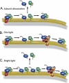
Mechanism of transducin translocation. A, the translocation of transducin requires the activation and separation of its functional subunits. The heterotrimer is tightly associated with the membrane as a result of the cooperative action of its two lipid moieties. Individual subunits have a lower membrane affinity, allowing their dissociation from the disc membranes while transducin remains activated. B, in dim light, activated transducin binds to PDE and is rapidly deactivated by the RGS9 complex before it can dissociate from the membrane. C, in bright light, translocation occurs when there is more activated transducin than PDE. This excess transducin, neither retained on the membrane by PDE nor rapidly deactivated by RGS9, stays activated sufficiently long to dissociate from the membrane to the cytosol and ultimately diffuse out of the rod outer segment. This figure was modified from Ref. with permission.
FIGURE 4.
FIGURE 4.
Faster arrestin-independent R* deactivation in cones than in rods. A, comparison of average normalized dim flash responses of WT and arrestin-1 knock-out (Arr1 KO) rods. The final response recovery in the arrestin knock-out rod has a time constant comparable with that for the thermal decay of R*, ∼50 s. B, comparison of average normalized dim flash responses of a WT cone and a double knock-out cone lacking both arrestin-1 and arrestin-4 (Arr1/Arr4 KO). The recovery phases of the cone responses are much more similar with and without arrestins; the double knock-out response decays with a final time constant of ∼750 ms. Traces in A were adapted from Ref. , and those in B from Ref. with permission.

References

    1. Burns M. E., Baylor D. A. (2001) Annu. Rev. Neurosci. 24, 779–805 - PubMed
    1. Fain G. L., Matthews H. R., Cornwall M. C., Koutalos Y. (2001) Physiol. Rev. 81, 117–151 - PubMed
    1. Arshavsky V. Y., Lamb T. D., Pugh E. N., Jr. (2002) Annu. Rev. Physiol. 64, 153–187 - PubMed
    1. Fu Y., Yau K. W. (2007) Pflugers Arch. 454, 805–819 - PMC - PubMed
    1. Maeda T., Imanishi Y., Palczewski K. (2003) Prog. Retin. Eye Res. 22, 417–434 - PubMed

Publication types

LinkOut - more resources