. 2011 Dec;5(6):517-26.
doi: 10.1016/j.molonc.2011.10.004. Epub 2011 Oct 20.

Focal adhesion kinase inhibitors are potent anti-angiogenic agents

Affiliations

Focal adhesion kinase inhibitors are potent anti-angiogenic agents

Miguel A Cabrita et al. Mol Oncol. 2011 Dec.

Abstract

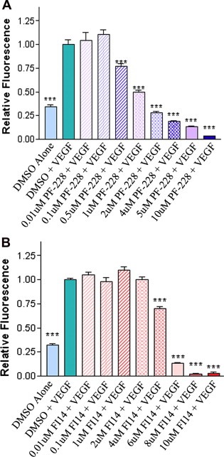
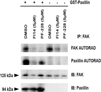
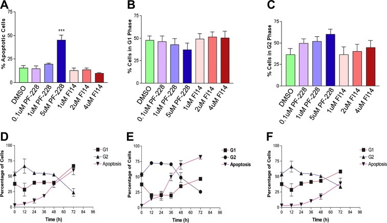
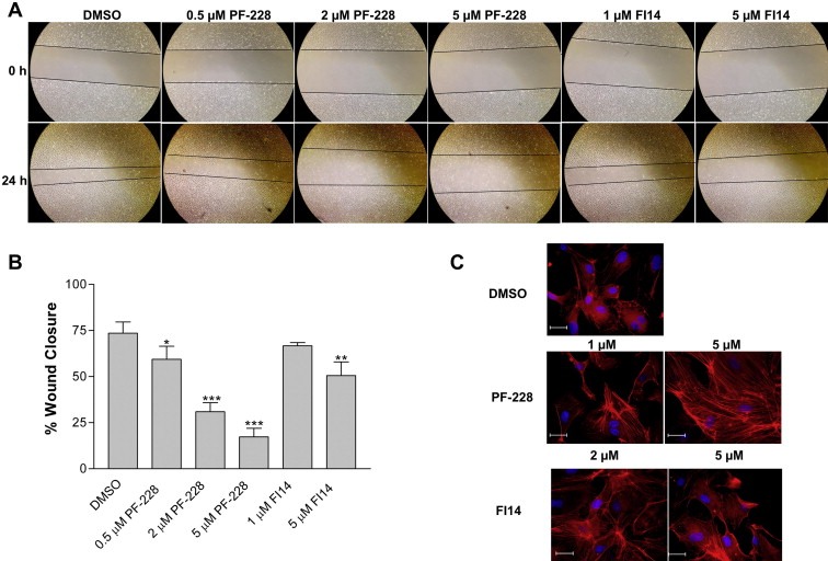
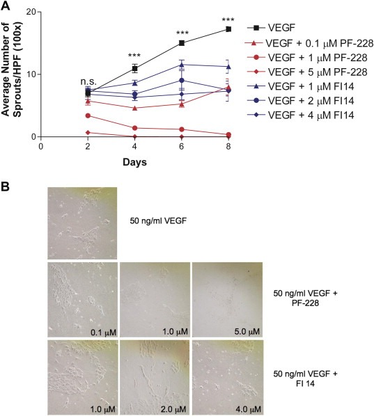
Focal adhesion kinase (FAK), a cytoplasmic tyrosine kinase and scaffold protein localized to focal adhesions, is uniquely positioned at the convergence point of integrin and receptor tyrosine kinase signal transduction pathways. FAK is overexpressed in many tumor cells, hence various inhibitors targeting its activity have been tested for anti-tumor activity. However, the direct effects of these pharmacologic agents on the endothelial cells of the vasculature have not been examined. Using primary human umbilical vein endothelial cells (HUVEC), we characterized the effects of two FAK inhibitors, PF-573,228 and FAK Inhibitor 14 on essential processes for angiogenesis, such as migration, proliferation, viability and endothelial cell tube formation. We observed that treatment with either FAK Inhibitor 14 or PF-573,228 resulted in reduced HUVEC viability, migration and tube formation in response to vascular endothelial growth factor (VEGF). Furthermore, we found that PF-573,228 had the added ability to induce apoptosis of endothelial cells within 36 h post-drug administration even in the continued presence of VEGF stimulation. FAK inhibitors also resulted in modification of the actin cytoskeleton within HUVEC, with observed increased stress fiber formation in the presence of drug. Given that endothelial cells were sensitive to FAK inhibitors at concentrations well below those reported to inhibit tumor cell migration, we confirmed their ability to inhibit endothelial-derived FAK autophosphorylation and FAK-mediated phosphorylation of recombinant paxillin at these doses. Taken together, our data indicate that small molecule inhibitors of FAK are potent anti-angiogenic agents and suggest their utility in combinatorial therapeutic approaches targeting tumor angiogenesis.


Figures

Figure 1
Figure 1
FAK inhibitors decrease number of viable endothelial cells in a dose‐dependent manner. HUVEC were incubated with various concentrations of either PF‐228 (A) or FI14 (B) in the presence of VEGF. Cells treated with vehicle (DMSO) were used as controls. Media containing alamarBlue was added 72 h post drug addition, to assess number of viable cells. Fluorescence was measured 6 h post‐alamarBlue addition. HUVEC treated with increasing concentrations of PF‐228 (A) or FI14 (B) showed reduced cell viability compared to DMSO controls. Experiments shown are representative of three or more experiments done in replicates of 8 wells per condition. Asterisks denote statistically significant differences (***p < 0.001) as determined by unpaired Student's t test.
Figure 2
Figure 2
FAK inhibitors FI14 and PF‐228 inhibit FAK kinase activity in vitro. FAK was immunoprecipitated from HUVEC total protein lysates and in vitro kinase assays were performed as described in Section 2.4, in the presence or absence of exogenously added recombinant paxillin as a kinase target substrate. Membranes were exposed to film to reveal the autoradiographic signals for phospho‐FAK and phospho‐paxillin (indicated by autorad panels). Membranes were subsequently analyzed by western blot for total FAK and paxillin as a loading control (indicated by IB panels). The amount of FAK autophosphorylation and paxillin target substrate phosphorylation by FAK was reduced by the presence of FAK inhibitors compared to vehicle control.
Figure 3
Figure 3
FAK inhibitors induce cell cycle arrest and apoptosis in treated endothelial cells. Seeded HUVEC were treated with DMSO as vehicle control or varying concentrations of PF‐228 or FI14 in the presence of 50 ng/mL VEGF for 48 h. Cells were then harvested and analyzed by flow cytometry as described in Section 2.5. The percentage of cells undergoing apoptosis (A) was quantified by examining cells with less than 2N DNA content following flow cytometric analysis on the FL2 channel. DNA content was used to calculate percentage of cells in G1 (B) and G2 (C) as described in Section 2.5. For the time course experiment, seeded HUVEC were incubated with VEGF (50 ng/ml) and DMSO as a vehicle control (D), or VEGF (50 ng/ml) in the presence of 5 μM PF‐228 (E) or 4 μM FI 14 (F). Cells were harvested at 12, 24, 36, 48 and 72 h post treatment, stained with propidium iodide and analyzed by flow cytometry as described in Section 2.5. Experiments shown are pooled results from three independent experiments performed in duplicate. Asterisks denote statistically significant differences (*p < 0.05, ***p < 0.001) as determined by unpaired Student's t test.
Figure 4
Figure 4
FAK inhibitors impede endothelial cell migration and alter the cellular actin cytoskeleton. HUVEC monolayers were wounded as described in Section 2.6, and cells were subsequently incubated with various concentrations of either PF‐228, FI14 or vehicle control (DMSO). Images were acquired at time of wounding (0 h) and 24 h post‐wounding (A, with lines indicating wound fronts) and percent wound closure was measured (B). Asterisks denote statistically significant differences (*p < 0.05, **p < 0.01, ***p < 0.001) as determined by unpaired Student's t test. HUVEC were grown on coverslips and treated with FAK inhibitors at the indicated concentrations for 24 h, prior to being stained as described in Section 2.7 with TRITC‐labeled phalloidin (red) to visualize actin filaments, and bisBenzimide Hoechst 33258 (blue) to visualize nuclei (C). More condensed phalloidin positive actin filaments were visualized in cells incubated with FAK inhibitors suggesting increased stress fiber formation. Experiments shown are representative of two independently performed experiments. Scale bar, 200 μM.
Figure 5
Figure 5
FAK inhibitors block HUVEC sprouting on collagen I gels. For assessing formation of sprouts, HUVEC were seeded onto collagen I gel‐coated plates and treated with 50 ng/ml VEGF in the absence (with added DMSO as a vehicle control) or presence of increasing concentrations of FAK inhibitors. HUVEC sprouting assay as a function of time (A). Sprouts were counted every 2 days in 10 random fields of view at 100× magnification. Mean number of sprouts per high powered field (HPF) and its associated standard error from duplicate dishes in each of two independent experiments are shown over time. Asterisks denote statistically significant differences (***p < 0.001) as determined by one‐way ANOVA analysis between cells treated with each of the drugs and vehicle. Sample images from day 8 of a representative experiment are shown (B). The effect of FAK inhibitors on sprout formation on collagen I was dose‐dependent.

References

    1. Abedi, H. , Zachary, I. , 1997. Vascular endothelial growth factor stimulates tyrosine phosphorylation and recruitment to new focal adhesions of focal adhesion kinase and paxillin in endothelial cells. The Journal of Biological Chemistry. 272, 15442–15451. - PubMed
    1. Abu-Ghazaleh, R. , Kabir, J. , Jia, H. , Lobo, M. , Zachary, I. , 2001. Src mediates stimulation by vascular endothelial growth factor of the phosphorylation of focal adhesion kinase at tyrosine 861, and migration and anti-apoptosis in endothelial cells. The Biochemical Journal. 360, 255–264. - PMC - PubMed
    1. Addison, C.L. , Nor, J.E. , Zhao, H. , Linn, S.A. , Polverini, P.J. , Delaney, C.E. , 2005. The response of VEGF-stimulated endothelial cells to angiostatic molecules is substrate-dependent. BMC Cell Biology. 6, 38 - PMC - PubMed
    1. Bagi, C.M. , Christensen, J. , Cohen, D.P. , Roberts, W.G. , Wilkie, D. , Swanson, T. , Tuthill, T. , Andresen, C.J. , 2009. Sunitinib and PF-562,271 (FAK/Pyk2 inhibitor) effectively block growth and recovery of human hepatocellular carcinoma in a rat xenograft model. Cancer Biology & Therapy. 8, 856–865. - PubMed
    1. Bagi, C.M. , Roberts, G.W. , Andresen, C.J. , 2008. Dual focal adhesion kinase/Pyk2 inhibitor has positive effects on bone tumors: implications for bone metastases. Cancer. 112, 2313–2321. - PubMed

Publication types

MeSH terms

Substances