

Review
doi: 10.1097/FJC.0b013e31821c0220.
A-kinase anchoring proteins that regulate cardiac remodeling
Affiliations
- PMID: 22075671
- PMCID: PMC3475412
- DOI: 10.1097/FJC.0b013e31821c0220
Item in Clipboard
Review
A-kinase anchoring proteins that regulate cardiac remodeling
J Cardiovasc Pharmacol.
2011 Nov.
Abstract
In response to injury or stress, the adult heart undergoes maladaptive changes, collectively defined as pathological cardiac remodeling. Here, we focus on the role of A-kinase anchoring proteins (AKAPs) in 3 main areas associated with cardiac remodeling and the progression of heart failure: excitation-contraction coupling, sarcomeric regulation, and induction of pathological hypertrophy. AKAPs are a diverse family of scaffold proteins that form multiprotein complexes, integrating cAMP signaling with protein kinases, phosphatases, and other effector proteins. Many AKAPs have been characterized in the heart, where they play a critical role in modulating cardiac function.
Figures
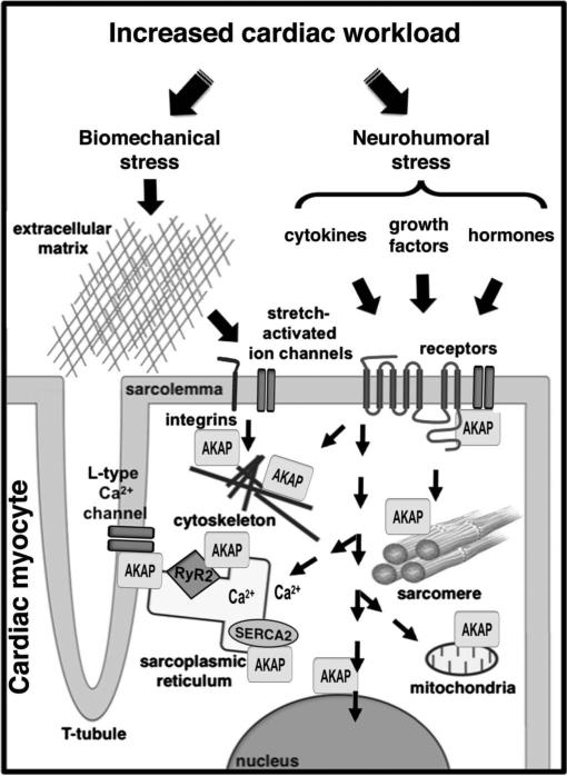
AKAPs coordinate signaling complexes in cardiac myocytes that function in cardiac remodeling. The adult heart responds to injury or stress by activating a variety of intracellular signaling pathways that may affect calcium handling, the cytoskeleton, and sarcomeric and mitochondrial function. Different AKAPs in cardiac myocytes play a critical role in coordinating these signaling events to promote reexpression of an embryonic gene program, myocyte hypertrophy, and extracellular matrix remodeling. Refer to the text for specific details.
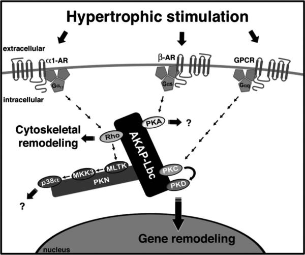
AKAP-Lbc coordinates hypertrophic signaling to promote cytoskeletal and gene remodeling. AKAP-Lbc is present in the cytoplasm, displaying a cytoskeletal and perinuclear localization. This anchoring protein serves as a scaffold for PKA, PKD, and its upstream activating kinase PKC. By bringing PKC and PKD into close proximity, AKAP-Lbc facilitates the phosphorylation and subsequent activation of PKD by PKC. Upon activation, PKD translocates to the nucleus promoting hypertrophic gene expression through phosphorylation of a histone deacetylase (HDAC5), leading to HDAC5 nuclear export and derepression of Mef2 transcription.AKAP-Lbc is a GEF for Rho. The Rho-GEF activity of AKAP-Lbc is stimulated via the Gα12 family of heterotrimeric G-proteins in response to α1-adrenergic receptor (AR) activation. GEF activity can be inactivated by an AKAP-Lbc anchored PKA-dependent mechanism. AKAP-Lbc also coordinates a p38α MAPK complex, downstream of Rho, composed of PKNα, MLTK, MKK3, and p38α. Presently, the downstream targets and functional consequences of this AKAP-Lbc-associated signaling cascade are unknown.
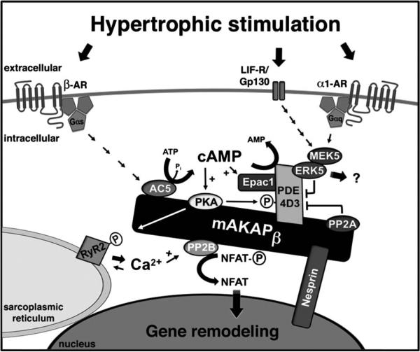
mAKAPβ coordinates hypertrophic signaling leading to cardiac gene remodeling. mAKAPβ is predominantly localized at the cardiomyocyte outer nuclear membrane through interaction with nesprin-1α. β-AR stimulation will result in the production of cAMP through Gs-coupled activation of mAKAPβ-bound adenylyl cyclase (AC5), likely present on transverse tubules adjacent to the nucleus. cAMP synthesis promotes mAKAP-bound PKA activation, leading to the phosphorylation of multiple substrates, including the RyR2, acting to potentiate Ca2+-induced RyR2 Ca2+ release. Local Ca2+ may activate mAKAP-associated PP2B (calcineurin Aβ), which will dephosphorylate the transcription factor NFATc, resulting in NFATc nuclear translocation and hypertrophic gene expression. cAMP metabolism is tightly regulated by the mAKAP signaling complex. PKA phosphorylates both AC5 and PDE4D3, inhibiting local cAMP production and increasing cAMP degradation, respectively. PKA also phosphorylates and stimulates PP2A, opposing PKA phosphorylation of PDE4D3, leading to greater, longer lasting cAMP signals. ERK5 will also phosphorylate and inhibit PDE4D3, thereby promoting PKA activity. α1-Adrenergic and gp130/leukemia inhibitory factor receptor (LIF-R) stimulation will result in MEK5 and ERK5 activation through a JAK/STAT/Ras/Raf pathway. Other substrates for mAKAPβ-associated ERK5 have not yet been identified. For simplicity, only a single mAKAP molecule is depicted.
References
-
- Frey N, Olson EN. Cardiac hypertrophy: the good, the bad, and the ugly. Annu Rev Physiol. 2003;65:45–79. - PubMed
-
- Heineke J, Molkentin JD. Regulation of cardiac hypertrophy by intracellular signalling pathways. NatRev MolCellBiol. 2006;7:589–600. - PubMed
-
- Chien KR. Stress pathways and heart failure. Cell. 1999;98:555–558. - PubMed
-
- Hill JA, Olson EN. Cardiac plasticity. N Engl J Med. 2008;358:1370–1380. - PubMed
-
- Barry SP, Townsend PA. What causes a broken heart—molecular insights into heart failure. Int Rev Cell Mol Biol. 2010;284:113–179. - PubMed
Publication types
MeSH terms
Substances
Grants and funding
LinkOut - more resources
Full Text Sources
