. 2012 Feb 1;302(3):C555-65.
doi: 10.1152/ajpcell.00299.2011. Epub 2011 Nov 9.

Thin-filament length correlates with fiber type in human skeletal muscle

Affiliations

Thin-filament length correlates with fiber type in human skeletal muscle

David S Gokhin et al. Am J Physiol Cell Physiol. .

Abstract

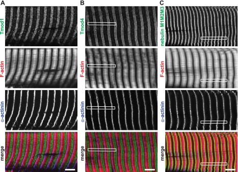
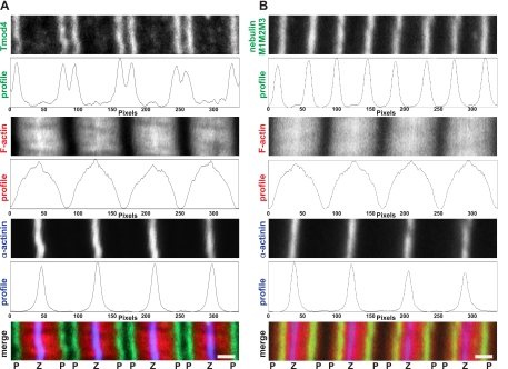
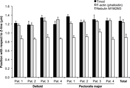
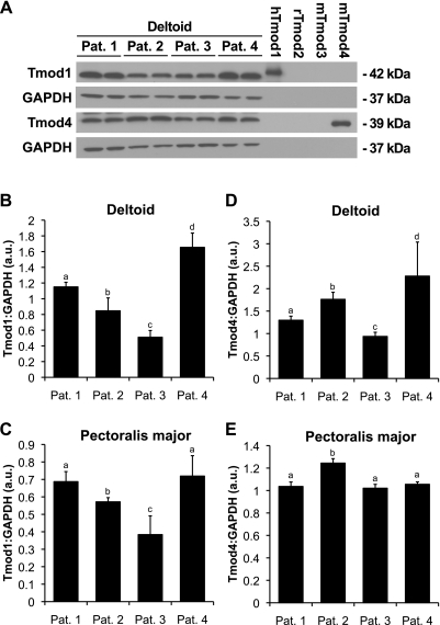
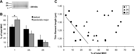
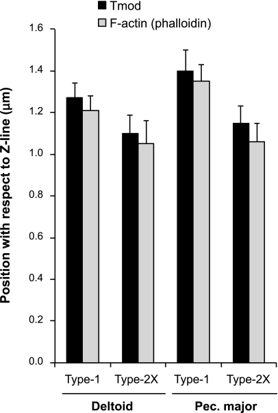
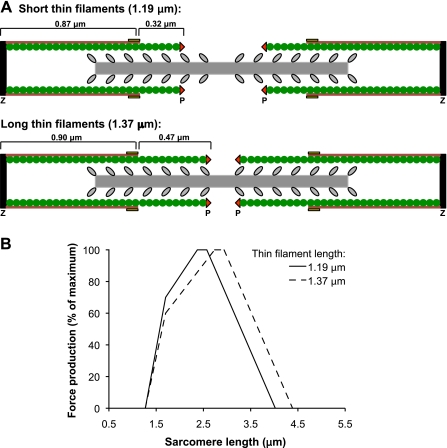
Force production in skeletal muscle is proportional to the amount of overlap between the thin and thick filaments, which, in turn, depends on their lengths. Both thin- and thick-filament lengths are precisely regulated and uniform within a myofibril. While thick-filament lengths are essentially constant across muscles and species (∼1.65 μm), thin-filament lengths are highly variable both across species and across muscles of a single species. Here, we used a high-resolution immunofluorescence and image analysis technique (distributed deconvolution) to directly test the hypothesis that thin-filament lengths vary across human muscles. Using deltoid and pectoralis major muscle biopsies, we identified thin-filament lengths that ranged from 1.19 ± 0.08 to 1.37 ± 0.04 μm, based on tropomodulin localization with respect to the Z-line. Tropomodulin localized from 0.28 to 0.47 μm further from the Z-line than the NH(2)-terminus of nebulin in the various biopsies, indicating that human thin filaments have nebulin-free, pointed-end extensions that comprise up to 34% of total thin-filament length. Furthermore, thin-filament length was negatively correlated with the percentage of type 2X myosin heavy chain within the biopsy and shorter in type 2X myosin heavy chain-positive fibers, establishing the existence of a relationship between thin-filament lengths and fiber types in human muscle. Together, these data challenge the widely held assumption that human thin-filament lengths are constant. Our results also have broad relevance to musculoskeletal modeling, surgical reattachment of muscles, and orthopedic rehabilitation.


Figures

Fig. 1.
Fig. 1.
Confocal fluorescence microscopy of human skeletal muscle thin-filament components. Shown are examples of longitudinal cryosections of pectoralis major muscle from patient 1 that were immunostained for tropomodulin (Tmod) 1 (A), Tmod4 (B), or the nebulin M1M2M3 domain (C), phalloidin stained for F-actin, and immunostained for α-actinin to visualize Z-lines. White rectangles indicate myofibrils that are magnified in Fig. 2. Bars, 5 μm.
Fig. 2.
Fig. 2.
Line-scan analysis of fluorescence intensity of thin-filament components during distributed deconvolution (DDecon) analysis. Higher magnification views of the boxed myofibrils in Fig. 1 are shown. Longitudinal cryosections of pectoralis major muscle from patient 1 were immunostained for Tmod4 (A) or the nebulin M1M2M3 domain (B), phalloidin stained for F-actin, and immunostained for α-actinin to visualize Z-lines. Each probe's corresponding fluorescence intensity profile used for DDecon analysis is shown below each myofibril. Note that the nebulin M1M2M3 domain is localized more proximally with respect to the Z-line compared with Tmod4. Z, Z-line; P, thin-filament pointed end. Bars, 1 μm.
Fig. 3.
Fig. 3.
Thin-filament lengths as determined by Tmod localization and F-actin breadth and the position of the NH2-terminal M1M2M3 domain of nebulin. Data are shown for individual biopsies and as pooled data. Note the more proximal position of the nebulin M1M2M3 domain with respect to the Z-line compared with Tmod. Error bars reflect means ± SD of myofibril counts shown in Table 2. Pat., patient.
Fig. 4.
Fig. 4.
Human skeletal muscles coexpress Tmod1 and Tmod4. A: Western blots of muscle tissue lysates probed with antibodies against Tmod1 and Tmod4. GAPDH was used for normalization. Note that native human Tmod1 (hTmod1) and Tmod4 migrate slightly lower and higher than purified recombinant hTmod1 and mouse Tmod4 (mTmod), respectively, which were used to verify the specificity of our Tmod isoform-specific antibodies. 10 ng of each Tmod1–4 were loaded on gels. rTmod2, rat Tmod2. BE: quantitation of Tmod1 (B and C) and Tmod4 (D and E) protein levels in the deltoid (B and D) and pectoralis major (C and E) muscles from each patient, with normalization to GAPDH. Error bars reflect means ± SD of n = 3 replicates. a,b,c,d | Different letters above bars within a single graph reflect significantly different values, as determined by one-way ANOVA with post hoc Fishers paired least significant difference tests. a.u., Arbitrary units.
Fig. 5.
Fig. 5.
Correlation between thin-filament length and myosin heavy chain (MHC) isoform composition in human skeletal muscles. A: muscle tissue lysates were separated on 8% SDS-PAGE gels and silver-stained. A sample separation of pectoralis major muscle from patient 1 is shown, and the positions of type 1, 2A, and 2X MHC are labeled. B: MHC isoform distributions. Error bars reflect means ± SD of n = 4 muscles. *P < 0.05, as determined by Student's t-test. C: correlation between thin-filament length (based on Tmod localization) and the percentages of type 1, 2A, and 2X MHC. Note that the proportion of type 2X MHC (R2 = 0.56; P < 0.01), but not type 1 (R2 = 0.14; P > 0.5) or type 2A (R2 = 0.07; P > 0.5) MHC is significantly correlated with thin-filament length.
Fig. 6.
Fig. 6.
Average thin-filament lengths in type 1 and type 2X MHC-positive fibers in deltoid and pectoralis major muscles in patient 1, as determined by Tmod localization and F-actin breadth. Note longer thin filaments in type 1 MHC-positive fibers and shorter thin filaments in type 2X MHC-positive fibers. Error bars reflect means ± SD of myofibril counts shown in Table 3.
Fig. <underline>7</underline>.
Fig. 7.
Hypothetical sarcomere length-tension curves of human skeletal muscles. A: model diagrams of sarcomeres with the minimum and maximum biopsy-wide thin-filament lengths measured in this study (1.19 and 1.37 μm, respectively) depict constant-length, nebulin-coated, thin-filament cores and variable-length, nebulin-free, pointed-end extensions that are capped by Tmod. Note that, for clarity, the sarcomere lengths are equal. However, at resting sarcomere lengths in vivo, the thin-filament pointed ends coincide with the thick-filament bare zone (38). Green, actin; red, Tmod; purple, nebulin; yellow, nebulin M1M2M3 domain; black, Z-line; gray, thick filament. B: length-tension curves were constructed based on thin-filament lengths of 1.19 and 1.37 μm, a constant thick-filament length of 1.65 μm, and a bare zone width of 0.2 μm (, –19, 24, 56).

References

    1. Almenar-Queralt A, Lee A, Conley CA, Ribas de Pouplana L, Fowler VM. Identification of a novel tropomodulin isoform, skeletal tropomodulin, that caps actin filament pointed ends in fast skeletal muscle. J Biol Chem 274: 28466–28475, 1999 - PubMed
    1. Bang ML, Li X, Littlefield R, Bremner S, Thor A, Knowlton KU, Lieber RL, Chen J. Nebulin-deficient mice exhibit shorter thin filament lengths and reduced contractile function in skeletal muscle. J Cell Biol 173: 905–916, 2006 - PMC - PubMed
    1. Broschat KO. Tropomyosin prevents depolymerization of actin filaments from the pointed end. J Biol Chem 265: 21323–21329, 1990 - PubMed
    1. Broschat KO, Weber A, Burgess DR. Tropomyosin stabilizes the pointed end of actin filaments by slowing depolymerization. Biochemistry 28: 8501–8506, 1989 - PubMed
    1. Carlier MF, Valentin-Ranc C, Combeau C, Fievez S, Pantoloni D. Actin polymerization: regulation by divalent metal ion and nucleotide binding, ATP hydrolysis and binding of myosin. Adv Exp Med Biol 358: 71–81, 1994 - PubMed

Publication types

MeSH terms

LinkOut - more resources