

Aging is associated with an increase in T cells and inflammatory macrophages in visceral adipose tissue
- PMID: 22075699
- PMCID: PMC3237772
- DOI: 10.4049/jimmunol.1102188
Aging is associated with an increase in T cells and inflammatory macrophages in visceral adipose tissue
Abstract
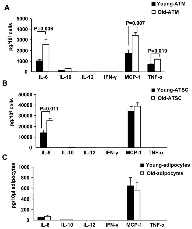
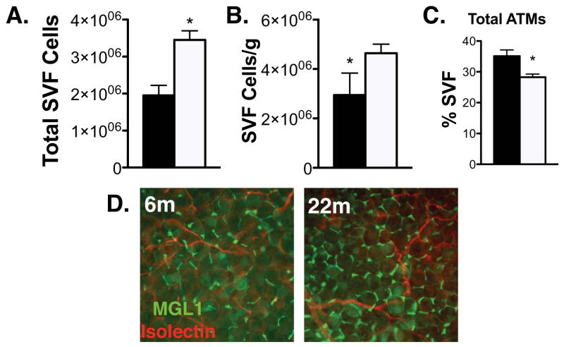
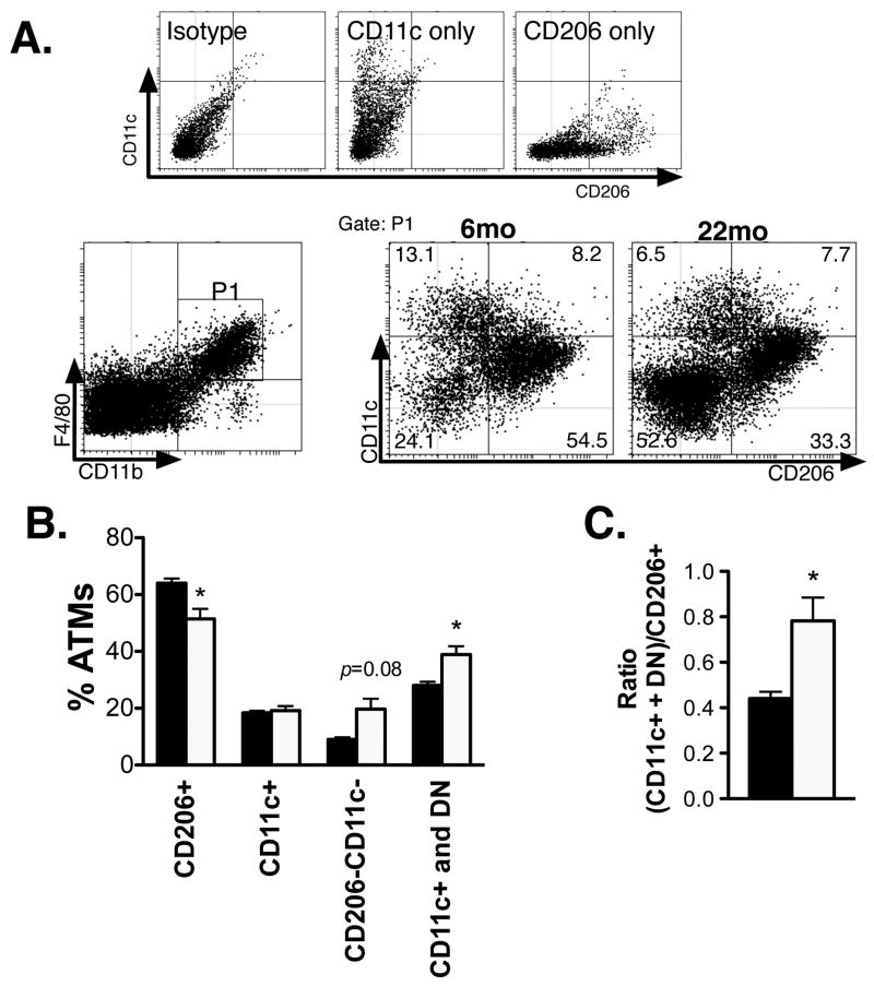
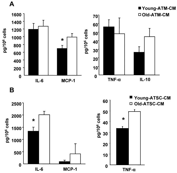
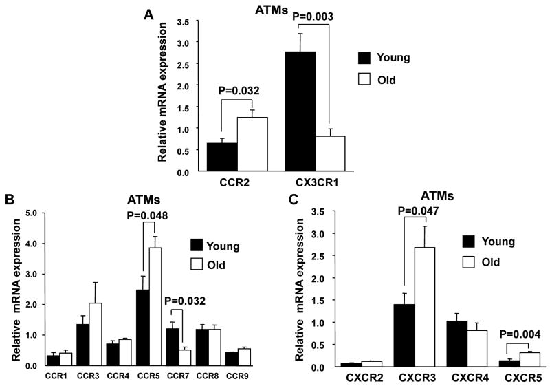
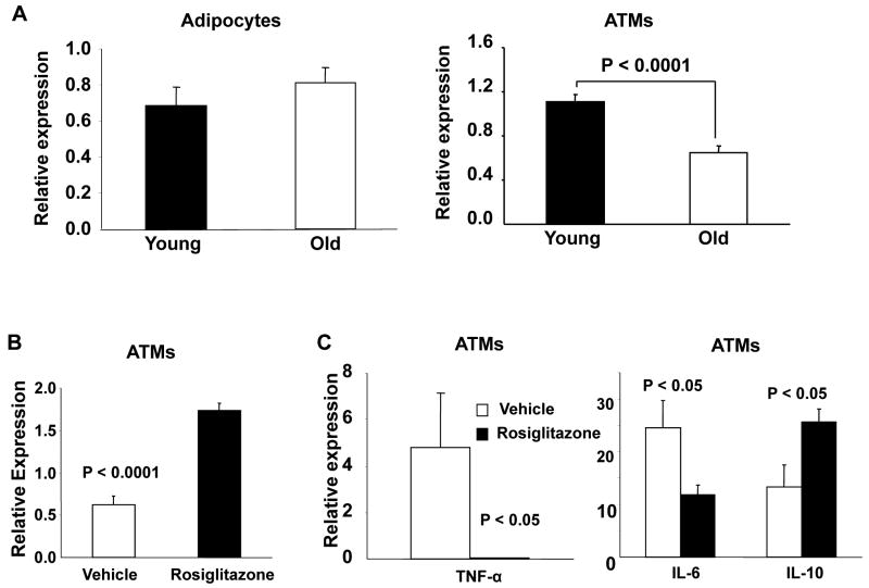
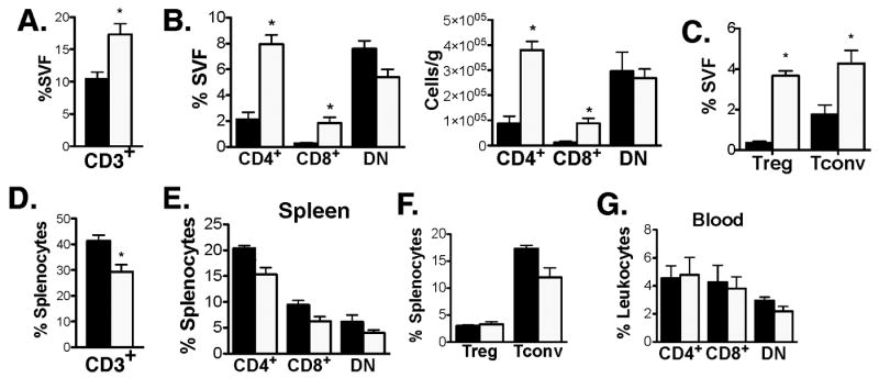
Age-related adiposity has been linked to chronic inflammatory diseases in late life. To date, the studies on adipose tissue leukocytes and aging have not taken into account the heterogeneity of adipose tissue macrophages (ATMs), nor have they examined how age impacts other leukocytes such as T cells in fat. Therefore, we have performed a detailed examination of ATM subtypes in young and old mice using state of the art techniques. Our results demonstrate qualitative changes in ATMs with aging that generate a decrease in resident type 2 (M2) ATMs. The profile of ATMs in old fat shifts toward a proinflammatory environment with increased numbers of CD206(-)CD11c(-) (double-negative) ATMs. The mechanism of this aging-induced shift in the phenotypic profile of ATMs was found to be related to a decrease in peroxisome proliferator-activated receptor-γ expression in ATMs and alterations in chemokine/chemokine receptor expression profiles. Furthermore, we have revealed a profound and unexpected expansion of adipose tissue T cells in visceral fat with aging that includes a significant induction of regulatory T cells in fat. Our findings demonstrate a unique inflammatory cell signature in the physiologic context of aging adipose tissue that differs from those induced in setting of diet-induced obesity.
Figures
References
-
- Leng SX, Yang H, Walston JD. Decreased cell proliferation and altered cytokine production in frail older adults. Aging Clin Exp Res. 2004;16:249–252. - PubMed
-
- Horber FF, Gruber B, Thomi F, Jensen EX, Jaeger P. Effect of sex and age on bone mass, body composition and fuel metabolism in humans. Nutrition. 1997;13:524–534. - PubMed
Publication types
MeSH terms
Grants and funding
- P30ES017885/ES/NIEHS NIH HHS/United States
- R01 AG020628/AG/NIA NIH HHS/United States
- R01AG020628/AG/NIA NIH HHS/United States
- R01DK090262/DK/NIDDK NIH HHS/United States
- P30 AG024824/AG/NIA NIH HHS/United States
- R01AG028268/AG/NIA NIH HHS/United States
- R01 DK090262/DK/NIDDK NIH HHS/United States
- P30 ES017885/ES/NIEHS NIH HHS/United States
- P30 AG013283/AG/NIA NIH HHS/United States
- R01 AG028268/AG/NIA NIH HHS/United States
- R01 AR042525/AR/NIAMS NIH HHS/United States
- P30 AR048310/AR/NIAMS NIH HHS/United States
- R56 AG020628/AG/NIA NIH HHS/United States
- P30AG024824/AG/NIA NIH HHS/United States
- R01AR042525/AR/NIAMS NIH HHS/United States
- K08 DK078851/DK/NIDDK NIH HHS/United States
- AG013283/AG/NIA NIH HHS/United States
- K08DK078851/DK/NIDDK NIH HHS/United States
- T32 GM007315/GM/NIGMS NIH HHS/United States
LinkOut - more resources
Full Text Sources
Other Literature Sources
Medical
Research Materials
Miscellaneous
