. 2011;6(11):e27213.
doi: 10.1371/journal.pone.0027213. Epub 2011 Nov 4.

Endogenous neurotrophins and Trk signaling in diffuse large B cell lymphoma cell lines are involved in sensitivity to rituximab-induced apoptosis

Affiliations

Endogenous neurotrophins and Trk signaling in diffuse large B cell lymphoma cell lines are involved in sensitivity to rituximab-induced apoptosis

Cynthia Bellanger et al. PLoS One. 2011.

Abstract

Background: Diffuse large B-cell lymphoma (DLBCL) is a common and often fatal malignancy. Immunochemotherapy, a combination of rituximab to standard chemotherapy, has resulted in improved survival. However a substantial proportion of patients still fail to reach sustained remission. We have previously demonstrated that autocrine brain-derived neurotrophic factor (BDNF) production plays a function in human B cell survival, at least partly via sortilin expression. As neurotrophin receptor (Trks) signaling involved activation of survival pathways that are inhibited by rituximab, we speculated that neurotrophins may provide additional support for tumour cell survival and therapeutic resistance in DLBCL.

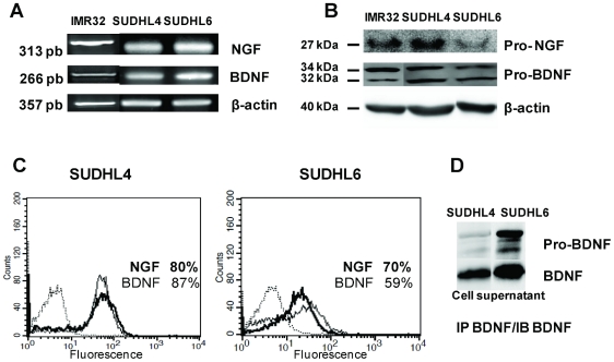
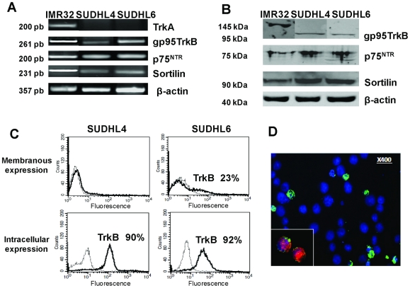
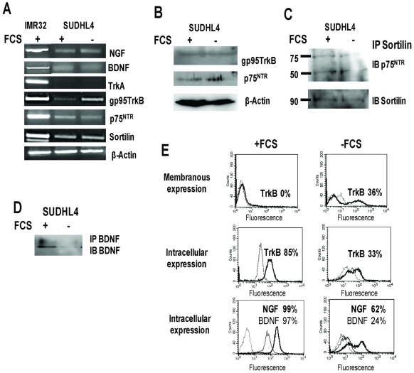
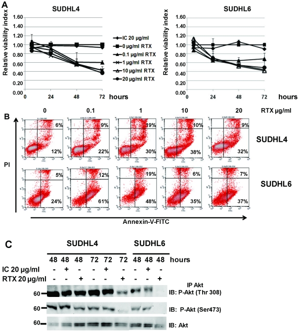
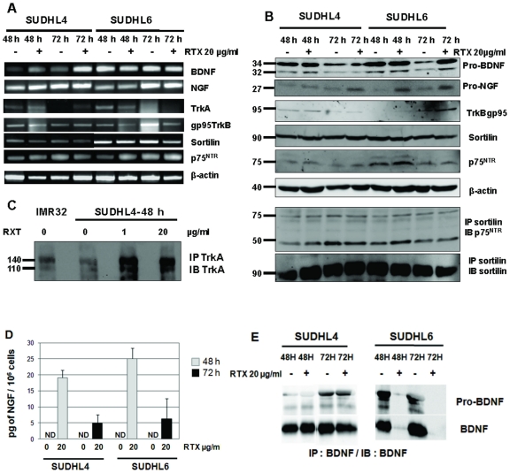
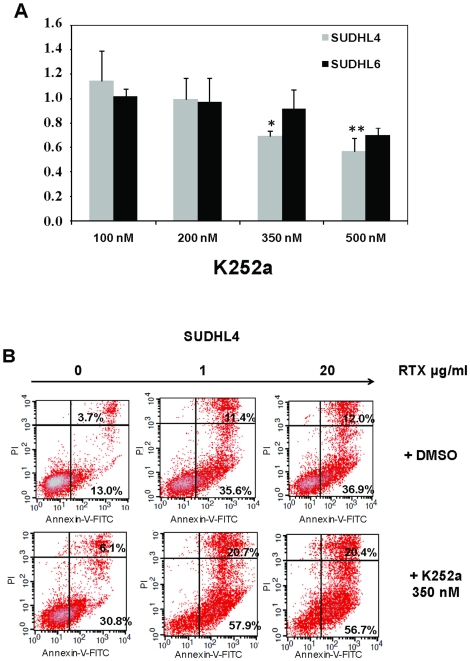
Methodology/principal findings: In the present study, we used two DLBCL cell lines, SUDHL4 and SUDHL6, known to be respectively less and more sensitive to rituximab. We found by RT-PCR, western blotting, cytometry and confocal microscopy that both cell lines expressed, in normal culture conditions, BDNF and to a lesser extent NGF, as well as truncated TrkB and p75(NTR)/sortilin death neurotrophin receptors. Furthermore, BDNF secretion was detected in cell supernatants. NGF and BDNF production and Trk receptor expression, including TrkA, are regulated by apoptotic conditions (serum deprivation or rituximab exposure). Indeed, we show for the first time that rituximab exposure of DLBCL cell lines induces NGF secretion and that differences in rituximab sensitivity are associated with differential expression patterns of neurotrophins and their receptors (TrkA). Finally, these cells are sensitive to the Trk-inhibitor, K252a, as shown by the induction of apoptosis. Furthermore, K252a exhibits additive cytotoxic effects with rituximab.

Conclusions/significance: Collectively, these data strongly suggest that a neurotrophin axis, such NGF/TrkA pathway, may contribute to malignant cell survival and rituximab resistance in DLBCL.


Conflict of interest statement

Competing Interests: The authors have declared that no competing interests exist.

Figures

Figure 1
Figure 1. DLBCL cell lines produce neurotrophins.
A: RT-PCR detection of NGF and BDNF mRNA was performed on SUDHL4 and SUDHL6 cells cultured with 10% FCS for 72 h. β-actin was included as a control of cDNA quality. B: Neurotrophin production was confirmed by western blotting of cell lysates demonstrating immature forms. Blots were reprobed with anti- β-actin as a loading control. C: Flow cytometry analysis demonstrating NGF (bold line) and BDNF intracellular expression in permeabilized cells (isotypic controls in dotted line). D: secretion of immature and mature BDNF was also detected in cell supernatants after immunoprecipitation (IP) and immunoblotting (IB) analysis. The neuroblastoma cell line, IMR32, was used as a positive control. Data are from one representative experiment out of four (RT-PCR, Flow cytometry and WB) or two (IP/WB) performed.
Figure 2
Figure 2. DLBCL cell lines express neurotrophin receptors.
A: RT-PCR detection of p75NTR, and its co-receptor sortilin, TrkA and truncated gp95TrkB mRNA was performed on SUDHL cells cultured with 10% FCS for 72 h. β-actin was included as a control of cDNA quality. B: The truncated TrkB, p75NTR and sortilin receptor protein expression was confirmed by western blotting of cell lysates. Blots were reprobed with anti- β-actin as a loading control. C: Flow cytometry analysis demonstrating TrkB membrane detection expressed in percentage of positive cells (bold line) in unpermeabilized SUDHL6 cells in contrast to SUDHL4 cells, whereas both cell lines expressed intracellular TrkB (dotted line: the isotypic controls). D: Immunofluorescence staining was performed, as described in Materials and Methods, in DLBCL cell lines that confirmed surface TrkB expression (in green) in some BDNF (inset in red) positive SUDHL6 cells (in blue: DAPI staining). The neuroblastoma cell line, IMR32, was used as a positive control. Data are representative of four independent experiments.
Figure 3
Figure 3. Modulation of neurotrophin and TrkB expression by SHDHL4 cells is induced by serum deprivation.
A: RT-PCR analysis of NGF, BDNF, TrkA, gp95TrkB, p75NTR and sortilin mRNA on SUDHL4 cells submitted to 72 h serum deprivation (−FCS) as compared to standard culture conditions (10% FCS, +FCS). β-actin was included as a control of cDNA quality. B: Western blott detection of truncated TrkB and p75NTR protein expression. Blots were reprobed with anti- β-actin as a loading control. C: Immunoprecipitation of sortilin (IP) and western blotting analysis (IB, immunoblotting with anti-p75NTR or anti-sortilin as control) demonstrating the association of sortilin with p75NTR in cell lysates of both culture conditions. D: secretion of BDNF detected in cell supernatants after immunoprecipitation and western blotting. E: Flow cytometry analysis demonstrating membranous expression of TrkB (bold line) and intracellular TrkB, NGF (bold line) and BDNF expressions expressed in percentage of SUDHL4 positive cells cultured with or without FCS (dotted line: isotypic controls). Data are representative of four independent experiments.
Figure 4
Figure 4. Effects of rituximab treatment on DLBCL cells lines.
A: Viability of SUDHL4 and SUDHL6 cells exposed to various concentrations of rituximab (RTX) during 72 h in serum basal culture condition (10% FCS). Viability was determined by XTT assay and calculated relative to time-matched untreated (0) controls. A control human IgG was also used as negative control (IC: Isotype control). Results are expressed as means ± SD of four experiments. B: Apoptosis induced by rituximab was measured after 24 h culture by Annexin-V-FITC/PI dual staining and the ratio of apoptotic (lower right quadrant) and necrotic (upper right quadrant) cells expressed as cell percentages was analyzed by flow cytometry. C: Rituximab induced inhibition of PI3K/Akt signaling which was detected by immunoprecipitation of Akt and western blott analysis of phosphorylated Akt (P-Akt) at 48 h and 72 h in SUDHL6 and SUDHL4 cell lysates. Data are representative of four independent experiments.
Figure 5
Figure 5. Effects of rituximab on neurotrophin production and Trk expression in DLBCL cells lines.
A: RT-PCR analysis of NGF, BDNF, TrkA, truncated TrkB, sortilin and p75NTR mRNA in SUDHL4 and SUDHL6 after 48 h and 72 h exposition to 20 µg/ml rituximab (RTX). β-actin was included as a control of cDNA quality. B: Western blott demonstrating in particular NGF protein expression following rituximab treatment and heterodimerization of p75NTR with sortilin in cell lysates. Blots were reprobed with anti- β-actin as a loading control. C: Enhanced TrkA expression induced by rituximab was observed for the less rituximab sensitive cell line, SUDHL4, after immunoprecipitation (IP) and immunoblotting (IB) with specific antibodies. D: NGF production was confirmed after rituximab exposure by detection with ELISA in cell supernatants of both cell lines, whereas it was undetectable (ND, non detected) in control cultures. Results are means ± SD of three independent experiments. E: BDNF secretion seemed to decrease in the most rituximab sensitive cell line, SUDHL6, as shown by western blott analysis of BDNF in cell lysate immunoprecipitates. Data are from one representative experiment out of four (RT-PCR, WB) or two (IP/WB) performed.
Figure 6
Figure 6. Pharmacologic inhibition of Trk receptors inhibits survival of DLBCL cell lines and synergizes rituximab induced apoptosis.
A: Cell viability was evaluated using the XTT test for both SUDHL cell lines cultured for 24 h in presence of various concentrations of K252a. Data are expressed as means ± SD of the relative cell viability of untreated control cells obtained from three independent experiments. Significant p values (*: p<0.05 and **: p<0.01) were determined in comparison with 100 nM K252a. B: K252a (350 nM) induced apoptosis in SUDHL4 cells, demonstrating a synergistic effect with rituximab (0–20 µg/ml) exposure. Example of flow cytometry analysis of apoptosis using Annexin-V-FITC and propidium iodide (PI), as detailed in legend of Figure 4, obtained after 48 h cell culture. Data are from one representative experiment out of three performed.

References

    1. De Paepe P, De Wolf-Peeters C. Diffuse large B-cell lymphoma: a heterogeneous group of non-Hodgkin lymphomas comprising several distinct clinicopathological entities. Leukemia. 2007;21:37–43. - PubMed
    1. Coiffier B, Pfreundschuh M, Stahel R, Vose J, Zinzani PL. Aggressive lymphoma: improving treatment outcome with rituximab. Anticancer Drugs. 2002;13:43–50. - PubMed
    1. Coiffier B. Rituximab therapy in malignant lymphoma. Oncogene. 2007;26:3603–3613. - PubMed
    1. Davis RE, Brown KD, Siebenlist U, Staudt LM. Constitutive nuclear factor κB activity is required for survival of activated B cell like diffuse large B cell lymphoma cells. J Exp Med. 2001;94:1861–1874. - PMC - PubMed
    1. Uddin S, Hussain AR, Siraj AK, Manogaran PS, Al-Jomah NA, et al. Role of phosphatidylinositol 3′-kinase/AKT pathway in diffuse large B-cell lymphoma survival. Blood. 2006;108:4178–4186. - PubMed

Publication types

MeSH terms