

Antidepressants stimulate hippocampal neurogenesis by inhibiting p21 expression in the subgranular zone of the hipppocampus
- PMID: 22076148
- PMCID: PMC3208633
- DOI: 10.1371/journal.pone.0027290
Antidepressants stimulate hippocampal neurogenesis by inhibiting p21 expression in the subgranular zone of the hipppocampus
Abstract
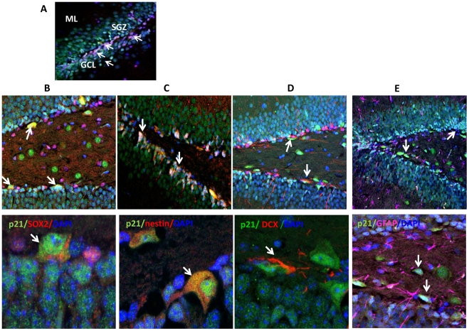
The relationships among hippocampal neurogenesis, depression and the mechanism of action of antidepressant drugs have generated a considerable amount of controversy. The cyclin-dependent kinase (Cdk) inhibitor p21(Cip1) (p21) plays a crucial role in restraining cellular proliferation and maintaining cellular quiescence. Using in vivo and in vitro approaches the present study shows that p21 is expressed in the subgranular zone of the dentate gyrus of the hippocampus in early neuronal progenitors and in immature neurons, but not in mature neurons or astroglia. In vitro, proliferation is higher in neuronal progenitor cells derived from p21-/- mice compared to cells derived from wild-type mice. Proliferation is increased in neuronal progenitor cells after suppression of p21 using lentivirus expressing short hairpin RNA against p21. In vivo, chronic treatment with the non-selective antidepressant imipramine as well as the norepinephrine-selective reuptake inhibitor desipramine or the serotonin-selective reuptake inhibitor fluoxetine all decrease p21 expression, and this was associated with increased neurogenesis. Chronic antidepressant treatment did not affect the expression of other Cdk inhibitors. Untreated p21-/- mice exhibit a higher degree of baseline neurogenesis and decreased immobility in the forced swim test. Although chronic imipramine treatment increased neurogenesis and reduced immobility in the forced swim test in wild-type mice, it reduced neurogenesis and increased immobility in p21-/- mice. These results demonstrate the unique role of p21 in the control of neurogenesis, and support the hypothesis that different classes of reuptake inhibitor-type antidepressant drugs all stimulate hippocampal neurogenesis by inhibiting p21 expression.
Conflict of interest statement
Figures


Similar articles
-
p21Cip1 restricts neuronal proliferation in the subgranular zone of the dentate gyrus of the hippocampus.Proc Natl Acad Sci U S A. 2008 Jan 29;105(4):1358-63. doi: 10.1073/pnas.0711030105. Epub 2008 Jan 2. Proc Natl Acad Sci U S A. 2008. PMID: 18172194 Free PMC article.
-
Antidepressants and Cdk inhibitors: releasing the brake on neurogenesis?Cell Cycle. 2008 Aug;7(15):2321-6. doi: 10.4161/cc.6446. Epub 2008 Jun 17. Cell Cycle. 2008. PMID: 18682686 Free PMC article. Review.
-
Hippocampal bone morphogenetic protein signaling mediates behavioral effects of antidepressant treatment.Mol Psychiatry. 2017 Jun;22(6):910-919. doi: 10.1038/mp.2016.160. Epub 2016 Oct 4. Mol Psychiatry. 2017. PMID: 27698430 Free PMC article.
-
Depression and adult neurogenesis: Positive effects of the antidepressant fluoxetine and of physical exercise.Brain Res Bull. 2018 Oct;143:181-193. doi: 10.1016/j.brainresbull.2018.09.002. Epub 2018 Sep 17. Brain Res Bull. 2018. PMID: 30236533 Review.
-
Neurogenic effects of fluoxetine are attenuated in p11 (S100A10) knockout mice.Biol Psychiatry. 2010 Jun 1;67(11):1048-56. doi: 10.1016/j.biopsych.2010.01.024. Epub 2010 Mar 15. Biol Psychiatry. 2010. PMID: 20227680
Cited by
-
Effects of Antidepressants on DSP4/CPT-Induced DNA Damage Response in Neuroblastoma SH-SY5Y Cells.Neurotox Res. 2015 Aug;28(2):154-70. doi: 10.1007/s12640-015-9534-z. Epub 2015 Jun 3. Neurotox Res. 2015. PMID: 26038195 Free PMC article.
-
Abnormal expression of cortical cell cycle regulators underlying anxiety and depressive-like behavior in mice exposed to chronic stress.Front Cell Neurosci. 2022 Dec 8;16:999303. doi: 10.3389/fncel.2022.999303. eCollection 2022. Front Cell Neurosci. 2022. PMID: 36568887 Free PMC article.
-
An Overview on Chemotherapy-induced Cognitive Impairment and Potential Role of Antidepressants.Curr Neuropharmacol. 2020;18(9):838-851. doi: 10.2174/1570159X18666200221113842. Curr Neuropharmacol. 2020. PMID: 32091339 Free PMC article. Review.
-
Corticosterone Impairs Hippocampal Neurogenesis and Behaviors through p21-Mediated ROS Accumulation.Biomolecules. 2024 Feb 23;14(3):268. doi: 10.3390/biom14030268. Biomolecules. 2024. PMID: 38540689 Free PMC article.
-
The reprotoxic adverse side effects of neurogenic and neuroprotective drugs: current use of human organoid modeling as a potential alternative to preclinical models.Front Pharmacol. 2024 Jun 14;15:1412188. doi: 10.3389/fphar.2024.1412188. eCollection 2024. Front Pharmacol. 2024. PMID: 38948466 Free PMC article. Review.
References
-
- Doetsch F. The glial identity of neural stem cells. Nat Neurosci. 2003;6:1127–1134. - PubMed
-
- Abrous DN, Koehl M, Le Moal M. Adult neurogenesis: from precursors to network and physiology. Physiol Rev. 2005;85:523–569. - PubMed
-
- Frazer A, Benmansour S. Delayed pharmacological effects of antidepressants. Mol Psychiatry. 2002;7(Suppl 1):S23–28. - PubMed
Publication types
MeSH terms
Substances
Grants and funding
LinkOut - more resources
Full Text Sources
Medical
Molecular Biology Databases
