. 2011;6(11):e27317.
doi: 10.1371/journal.pone.0027317. Epub 2011 Nov 4.

An in vivo polymicrobial biofilm wound infection model to study interspecies interactions

Affiliations

An in vivo polymicrobial biofilm wound infection model to study interspecies interactions

Trevor Dalton et al. PLoS One. 2011.

Abstract

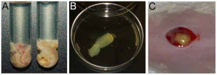
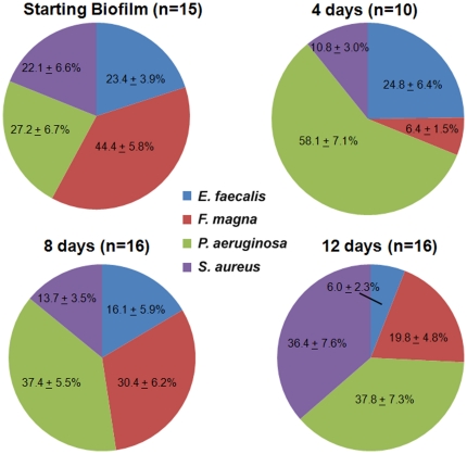
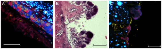
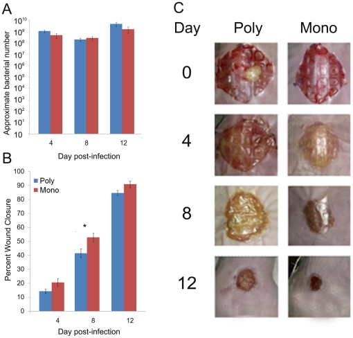
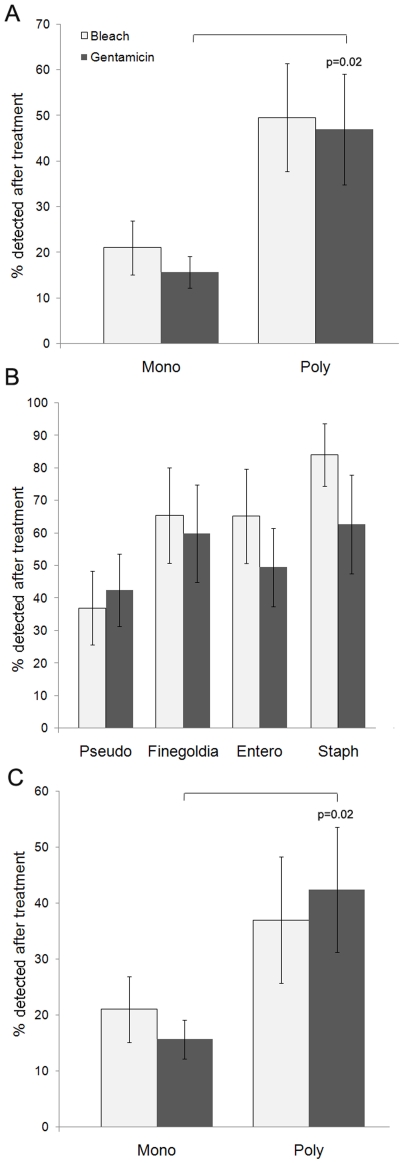
Chronic wound infections are typically polymicrobial; however, most in vivo studies have focused on monospecies infections. This project was designed to develop an in vivo, polymicrobial, biofilm-related, infected wound model in order to study multispecies biofilm dynamics and in relation to wound chronicity. Multispecies biofilms consisting of both Gram negative and Gram positive strains, as well as aerobes and anaerobes, were grown in vitro and then transplanted onto the wounds of mice. These in vitro-to-in vivo multi-species biofilm transplants generated polymicrobial wound infections, which remained heterogeneous with four bacterial species throughout the experiment. We observed that wounded mice given multispecies biofilm infections displayed a wound healing impairment over mice infected with a single-species of bacteria. In addition, the bacteria in the polymicrobial wound infections displayed increased antimicrobial tolerance in comparison to those in single species infections. These data suggest that synergistic interactions between different bacterial species in wounds may contribute to healing delays and/or antibiotic tolerance.


Conflict of interest statement

Competing Interests: RW and SD are affiliated with a commercial company (Research and Testing Laboratory, Lubbock, Texas). This does not alter the authors' adherence to all the PLoS ONE policies on sharing data and materials.

Figures

Figure 1
Figure 1. In vitro-to-in vivo polymicrobial biofilm transplant.
A. Biofilms were grown in vitro with four different bacterial species, as described in the text. A comparison between an in vitro grown biofilm (left) and an actual wound debridement from a wound patient (right) are shown to demonstrate the textural similarity between this specialized media and an actual wound. B. Mature biofilms were rinsed in sterile saline and sectioned. C. biofilm sections were seeded onto the wounds of mice.
Figure 2
Figure 2. Relative population distribution of starting in vitro-grown biofilms and 4, 8 and 12-day wound infections.
Realtime PCR was performed using species-specific primers with comparable amplification efficiencies in order to determine the relative ratio of the four different species. All mice that were infected with polymicrobial biofilms had detectable levels of all four organisms in their wounds. Average of groups ± SEM are shown.
Figure 3
Figure 3. Sections of in vitro-grown multispecies biofilms (A) or tissue from 12-day old infected murine wounds (B) were fixed in formalin, embedded in paraffin, thin-sectioned and stained with H&E.
Arrows indicate groups of morphologically distinct bacteria, scale = 10 µm.
Figure 4
Figure 4. Homogeneous ‘pockets’ of bacteria were visualized along the wound margin of 12-day-old infected wounds.
Wound tissue was fixed in formalin, embedded in paraffin, thin-sectioned and either stained with H&E (A) or hybridized to species-specific FISH probes (B), where P. aeruginosa is shown in red, S. aureus in yellow, and E. faecalis, F. magna, and host cell DNA are stained with DAPI (blue), scale = 10 µm.
Figure 5
Figure 5. P. aeruginosa makes up the leading edge of the infection.
(A) In vivo biofilms were imaged with FISH. Sections from the wound margins of 12-day-old infected mouse wounds were fixed in formalin, embedded in paraffin, thin-sectioned and mounted on slides. Sections were hybridized to a ‘target’ DNA probe complementary to a specific 16S region of the bacterial ribosomal subunit of either P. aeruginosa (red) or S. aureus (yellow) and stained with DAPI (E. faecalis, F. magna, and host cell DNA), scale = 50 µm. DAPI-stained host cell nuclei in the uninfected dermis are visible in the top right, followed by a polymicrobial-infected layer of the wound eschar, which is bordered by a layer of predominately P. aeruginosa extending into the wound bed. ‘Budding’ projections were visualized in the wound sections from 12-day-old polymicrobial infected mice by H&E (B) and FISH (C), scale = 10 µm. These projections extended from the leading edge of the wound margin, into the wound bed and hybridized to the P. aeruginosa 16S FISH probe (see in red, C).
Figure 6
Figure 6. The percent closure and bacterial load was determined for wounds infected polymicrobial biofilms or P. aeruginosa alone.
Realtime PCR analysis was used to approximate the bacterial number (A) in the infected tissue at 4, 8 and 12 days post-infection, n = 16 mice/time point. Percent wound closure (B) was determined at 4, 8 and 12 days post-infection and ANOVA with Tukey-Kramer Comparison's Test was used to determine statistical differences between groups, n = 16 mice/time point. There was no statistical difference in the bacterial load data. For the wound closure data, *p<0.05. Representative wound images are shown (C).
Figure 7
Figure 7. A higher percentage of bacterial cells from polymicrobial wound infections were detected after treatment with antimicrobials than those from monospecies (P. aeruginosa) infections (A).
Percentage of each bacterial species from polymicrobial wound infections that were detected after treatment with antimicrobials (B). Percentage of P. aeruginosa cells, either from monospecies or polymicrobial wound infections, which were detected after treatment with antimicrobials (C). The number of bacteria in treated and untreated samples was analyzed using realtime PCR, n = 6–8 mice/group. The Mann-Whitney Test was used to determine statistical differences between groups and the two-tailed p value is shown.

Similar articles

Cited by

References

    1. American Burn Association website. Burn Incidence and Treatment in the US: 2007 Fact Sheet. Available: http://www.ameriburn.org/resources_factsheet.php. Accessed 2011 Oct 17.
    1. Center for Disease Control. . Department of Health and Human Services, Centers for Disease Control and Prevention (Eds.) Atlanta, GA: 2002. National estimates and general information on diabetes in the U.S. Revised edition in:Center U.S.
    1. National Institute of General Medical Sciences website. Fact Sheet:Trauma, Shock, Burn and Injury: Fact and Figures. 2007;17 http://www.nigms.nih.gov/Education/Factsheet_Trauma.htm. Accessed 2011 Oct.
    1. James GA, Swogger E, Wolcott R, Pulcini E, Secor P, et al. Biofilms in chronic wounds. Wound Repair Regen. 2008;16:37–44. - PubMed
    1. Perencevich EN, Sands KE, Cosgrove SE, Guadagnoli E, Meara E, et al. Health and economic impact of surgical site infections diagnosed after hospital discharge. Emerg Infect Dis. 2003;9:196–203. - PMC - PubMed

Publication types

MeSH terms