Gastric lipomatosis
- PMID: 22076194
- PMCID: PMC3204505
- DOI: 10.5230/jgc.2010.10.4.254
Gastric lipomatosis
Abstract
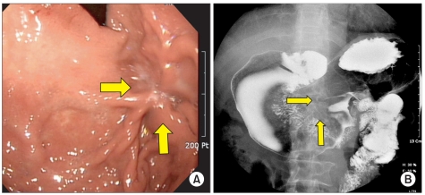
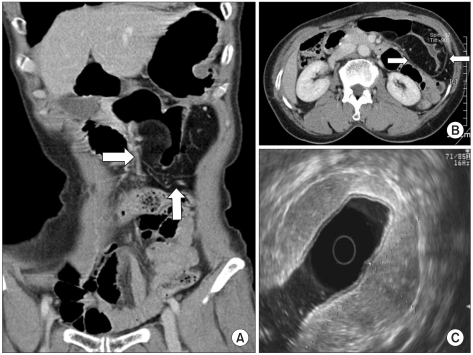
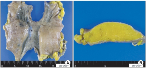
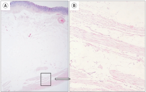
Gastric lipomatosis is an extremely rare condition. We present a case of a 69-year-old woman admitted with epigastric soreness. Computerized tomography (CT) revealed extrinsically compressing, fat-containing mass lesions on the entire gastric wall of the antrum and body except for the lesser curvature. A subtotal gastrectomy was performed. Pathology findings confirmed a gastric lipomatosis with multiple gastric ulcerations and extensive disruptions of the muscular layers. This case and reports of other gastric lipomatosis cases indicate that CT should be used to characterize large submucosal masses because CT can show the specific nature and extent of the disease. We believe that surgical treatment is the most appropriate treatment for symptomatic gastric lipomatosis that shows extensive gastric involvement, or when there are multiple gastric lipomas.
Keywords: Gastrectomy; Lipomatosis; Stomach neoplasms.
Figures
References
-
- Kim HS, Noh SH, Kim CK. Lipomas of gastrointestinal tract. J Korean Surg Soc. 1989;36:98–108.
-
- Ferrozzi F, Tognini G, Bova D, Pavone P. Lipomatous tumors of the stomach: CT findings and differential diagnosis. J Comput Assist Tomogr. 2000;24:854–858. - PubMed
-
- Suárez Moreno RM, Hernández Ramírez DA, Madrazo Navarro M, Salazar Lozano CR, Martínez Gen R. Multiple intestinal lipomatosis. Case report. Cir Cir. 2010;78:163–165. - PubMed
-
- Ventura L, Leocata P, Guadagni S, Ventura T. Multiple gastric lipomas: report of an asymptomatic case found at autopsy. Pathol Int. 1997;47:575–577. - PubMed
Publication types
LinkOut - more resources
Full Text Sources