. 2011 Nov 1;2(11):2934-49.
doi: 10.1364/BOE.2.002934. Epub 2011 Sep 30.

Model of bleaching and acquisition for superresolution microscopy controlled by a single wavelength

Affiliations

Model of bleaching and acquisition for superresolution microscopy controlled by a single wavelength

Alex Small. Biomed Opt Express. .

Abstract

We consider acquisition schemes that maximize the fraction of images that contain only a single activated molecule (as opposed to multiple activated molecules) in superresolution localization microscopy of fluorescent probes. During a superresolution localization microscopy experiment, irreversible photobleaching destroys fluorescent molecules, limiting the ability to monitor the dynamics of long-lived processes. Here we consider experiments controlled by a single wavelength, so that the bleaching and activation rates are coupled variables. We use variational techniques and kinetic models to demonstrate that this coupling of bleaching and activation leads to very different optimal control schemes, depending on the detailed kinetics of fluorophore activation and bleaching. Likewise, we show that the robustness of the acquisition scheme is strongly dependent on the detailed kinetics of activation and bleaching.

Keywords: (100.6640) Superresolution; (180.2520) Fluorescence microscopy.


Figures

Fig. 1
Fig. 1
Schematic of states and transitions for a fluorophore in which the dark state is the default state and bleaching occurs from the activated state. We assume multiple vibrational sublevels in the activated and excited states, to account for Stokes shifts of the absorption and emission spectra. The bleaching process depicted occurs from the excited state, and is assumed to not require the absorption of an additional molecule from the excited state.
Fig. 2
Fig. 2
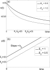
(a) n(t) and (b) p(t) for acquisition at different constant error rates, under scenario 1.
Fig. 3
Fig. 3
Schematic of a fluorophore whose default state is activated (i.e. fluorescent). Three plausible bleaching pathways are illustrated, numbered in the order in which they are considered here. Blue upward arrows indicate absorption of a photon, solid diagonal lines indicate bleaching upon the absorption of an additional photon, and diagonal dashed lines indicate bleaching without the absorption of an additional photon.
Fig. 4
Fig. 4
(a) n(t) and (b) p(t) for acquisition at constant error rate, under scenario 2. The time at which the number of molecules has decreased by half is shown for each plot in (a).
Fig. 5
Fig. 5
Solution to Eq. (38) for different initial error rates.
Fig. 6
Fig. 6
Excitation intensity I(t) for different initial error rates .

References

    1. Lidke K., Rieger B., Jovin T., Heintzmann R., “Superresolution by localization of quantum dots using blinking statistics,” Opt. Express 13, 7052–7062 (2005).10.1364/OPEX.13.007052 - DOI - PubMed
    1. Betzig E., Patterson G. H., Sougrat R., Lindwasser O. W., Olenych S., Bonifacino J. S., Davidson M. W., Lippincott-Schwartz J., Hess H. F., “Imaging intracellular fluorescent proteins at nanometer resolution,” Science 313, 1642–1645 (2006).10.1126/science.1127344 - DOI - PubMed
    1. Rust M. J., Bates M., Zhuang X., “Sub-diffraction-limit imaging by stochastic optical reconstruction microscopy (storm),” Nat. Methods 3, 793–795 (2006).10.1038/nmeth929 - DOI - PMC - PubMed
    1. Hess S. T., Girirajan T. P. K., Mason M. D., “Ultra-high resolution imaging by fluorescence photoactivation localization microscopy,” Biophys. J. 91, 4258–4272 (2006).10.1529/biophysj.106.091116 - DOI - PMC - PubMed
    1. Thompson R. E., Larson D. R., Webb W. W., “Precise nanometer localization analysis for individual fluorescent probes,” Biophys. J. 82, 2775–2783 (2002).10.1016/S0006-3495(02)75618-X - DOI - PMC - PubMed

LinkOut - more resources