. 2011 Nov 1;2(11):3129-34.
doi: 10.1364/BOE.2.003129. Epub 2011 Oct 25.

Swept source optical coherence tomography as a tool for real time visualization and localization of electrodes used in electrophysiological studies of brain in vivo

Swept source optical coherence tomography as a tool for real time visualization and localization of electrodes used in electrophysiological studies of brain in vivo

Hideyuki Watanabe et al. Biomed Opt Express. .

Abstract

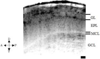
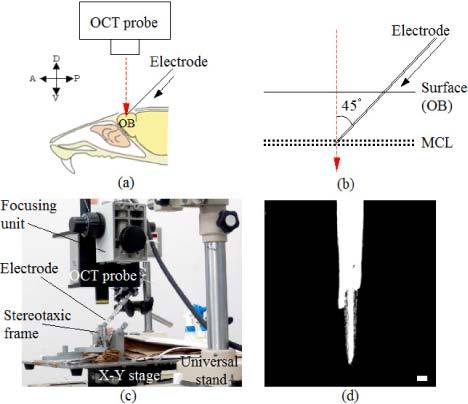
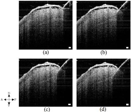
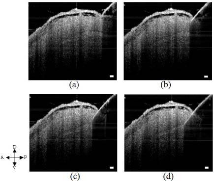
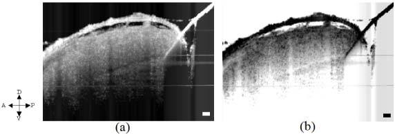
In studies of in vivo extracellular recording, we usually penetrate electrodes almost blindly into the neural tissue, in order to detect the neural activity from an expected target location at a certain depth. After the recording, it is necessary for us to determine the position of the electrodes precisely. Generally, to identify the position of the electrode, one method is to examine the postmortem tissue sample at micron resolution. The other method is using MRI and it does not have enough resolution to resolve the neural structures. To solve such problems, we propose swept source optical coherence tomography (SS-OCT) as a tool to visualize the cross-sectional image of the neural target structure along with the penetrating electrode. We focused on a rodent olfactory bulb (OB) as the target. We succeeded in imaging both the OB layer structure and the penetrating electrode, simultaneously. The method has the advantage of detecting the electrode shape and the position in real time, in vivo. These results indicate the possibility of using SS-OCT as a powerful tool for guiding the electrode into the target tissue precisely in real time and localizing the electrode tip during electrophysiological recordings.

Keywords: (170.3880) Medical and biological imaging; (170.4500) Optical coherence tomography; (170.5380) Physiology.


Figures

Fig. 1
Fig. 1
Neural layer structure of OB imaged by using SS-OCT. OB consists of distinctive different layers, namely: glomerulous layer (GL), external plexiform layer (EPL), mitral cell body layer (MCL) and granule cell layer (GCL). Here, we choose MCL as our specific target layer that is known to contain mainly cell bodies from which extracellular recordings are usually done. The arrows on the left corner indicate the anterior-posterior and dorsal-ventral parts of the rat. A, anterior; P, posterior; D, dorsal; V, ventral. Scale bar, 100 μm.
Fig. 2
Fig. 2
(a) A schematic view of the olfactory bulb (OB) with an electrode penetration and the OCT probe unit. The arrows on the left corner indicate the anterior-posterior and dorsal-ventral parts of the rat. A, anterior; P, posterior; D, dorsal; V, ventral. (b) A schematic of the magnified view of the electrode into mitral cell body layer (MCL) of OB. The angle between the optical axis and the electrode was set to be approximately 45 degree. (c) An optical micrograph of the optical probe unit of SS-OCT and the electrode probe unit. (d) An optical micrograph of the electrode with the external insulator seen as white and the exposed tungsten electrode seen rather as gray with the scale bar corresponding to 50 μm.
Fig. 3
Fig. 3
Raw OCT B-scan images obtained during the electrode penetration process at different times (a) t = 0, (b) t = 7, (c) t = 10.5, and (d) t = 14 sec. The penetration process was monitored and finally reaching the target location of MCL. We saw granular structure corresponding to speckles appearing as a result of multiple scattering within the probing coherence volume. The arrows on the left corner indicate the anterior-posterior and dorsal-ventral parts of the rat. A, anterior; P, posterior; D, dorsal; V, ventral. Scale bar, 100 μm. Refer to the movie (Media 1) of raw images.
Fig. 4
Fig. 4
Five-frame averaged OCT B-scan images obtained during the electrode penetration process at different times (a) t = 0 sec; (b) t = 7 sec; (c) t = 10.5 sec; (d) t = 14 sec. The penetration process was clearly seen and finally reaching the target location of MCL. The arrows on the left corner indicate the anterior-posterior and dorsal-ventral parts of the rat. A, anterior; P, posterior; D, dorsal; V, ventral. Scale bar, 100 μm. Refer to the movie (Media 2) of five-frame averaged images.
Fig. 5
Fig. 5
(a) Averaged OCT B-scan image of OB with the stationary electrode being positioned at the target location MCL of OB. (b) Averaged OCT image same as (a) with inverted dynamic range to have a clear visualization of the target layer in relation to the electrode. Part below the electrode in the OCT signal was not detectable because the electrode practically reflected the sample beam. The arrows on the left corner indicate the anterior-posterior and dorsal-ventral parts of the rat. A, anterior; P, posterior; D, dorsal; V, ventral. Scale bar, 100 μm.

Similar articles

Cited by

References

    1. Fantana A. L., Soucy E. R., Meister M., “Rat olfactory bulb mitral cells receive sparse glomerular inputs,” Neuron 59(5), 802–814 (2008).10.1016/j.neuron.2008.07.039 - DOI - PubMed
    1. Nagayama S., Takahashi Y. K., Yoshihara Y., Mori K., “Mitral and tufted cells differ in the decoding manner of odor maps in the rat olfactory bulb,” J. Neurophysiol. 91(6), 2532–2540 (2004).10.1152/jn.01266.2003 - DOI - PubMed
    1. Griff E. R., Mafhouz M., Perrut A., Chaput M. A., “Comparison of identified mitral and tufted cells in freely breathing rats: I. Conduction velocity and spontaneous activity,” Chem. Senses 33(9), 779–792 (2008).10.1093/chemse/bjn041 - DOI - PubMed
    1. Chapuis J., Garcia S., Messaoudi B., Thevenet M., Ferreira G., Gervais R., Ravel N., “The way an odor is experienced during aversive conditioning determines the extent of the network recruited during retrieval: a multisite electrophysiological study in rats,” J. Neurosci. 29(33), 10287–10298 (2009).10.1523/JNEUROSCI.0505-09.2009 - DOI - PMC - PubMed
    1. Sato T., Uchida G., Tanifuji M., “Cortical columnar organization is reconsidered in inferior temporal cortex,” Cereb. Cortex 19(8), 1870–1888 (2009).10.1093/cercor/bhn218 - DOI - PMC - PubMed

LinkOut - more resources